
In-solution characterization of affinity and structural properties of the FcRn IgG1 Fc interaction during product development
INTRODUCTION
The pharmacokinetics (PK) of therapeutic antibodies are typically influenced by multiple factors (e.g., administration route and dose) but antibody design can also have an impact on PK by, for example, modifying the Fc fragment sequence (e.g., YTE1) to increase affinity for FcRn (neonatal Fc receptor) to modulate the drug’s half-life in serum.
The identification of degradation pathways can additionally be benefited by FcRn affinity characterization. As a result, characterising FcRn interactions with therapeutic antibodies is a common practice in drug development and a surrogate means of assessing the possible influence on PK by molecule design or product stability.
FIDA was evaluated as an analytical approach for characterising the design of FcRn-related antibody-Fc alterations using three IgG1-Fc variants (IgG1-YTE, IgG1-TM2, and IgG1-YTE-TM) vs an IgG1 wild type (WT). The IgG1-YTE variant is utilised to alter the PK by extending the half-life of the therapeutic antibody in serum. However, the TM alteration is occasionally used separately or in concert with the YTE variation in the therapeutic design to abrogate the putative effector function activity. The FcRn affinity increase caused by the IgG1-YTE alteration is predicted to be unaffected by the TM addition, and the WT affinity is expected to be equivalent to the IgG1-TM variation because TM is not intended to extend or shorten the antibody’s half-life.
The FcRn receptor is known to play crucial roles in the neonatal transfer of antibodies across the placenta and the recycling of IgGs by binding with pH-dependent affinity which modulates the PK of mAb drugs. The binding of FcRn and Fc at a specific low pH is the main topic of interest in this application note.
We employed several analytical techniques to characterize the Fc-FcRn interaction. To quantify affinity (Kd), label-free protein-protein interaction technology (SPR) is often utilised. A relative quantitation in-solution immunoassay is also commonly employed to offer EC50 values rather than affinity constants.
Because of the influence on the technique’s readout, the latter method is not always appropriate for proteins having self-association properties. We provide a new quantitative technique to assessing the Fc-FcRn interaction utilising Flow-Induced Dispersion Analysis (FIDA measures the fluorescence of particles in laminar flow, analyses their dispersion, and calculates the hydrodynamic radius of particles) to produce affinity data from an in-solution technology.
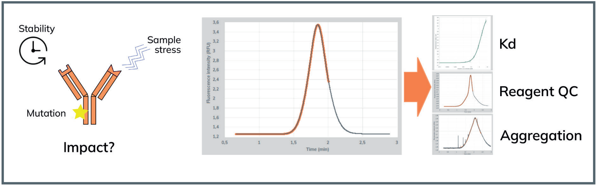
FIDA is capable of not only characterising the design of the Fc variants and identifying some Fc:FcRn associated product-specific quality attributes with functional data, but also of evaluating the quality of the reagents used during the experiment by identifying, for example, protein aggregation formation of the labelled reagent in a high-throughput Taylor grams.
MATERIALS AND METHODS
Fida 1 instrument equipped with a 480 nm LED fluorescence detector. A Fidabio dynamic coated capillary (L: 1 m, ID: 75 µm, Leff: 84 cm).
The buffer used was 50 mM NaPhos, 100 mM NaCl, 0.005% P20, pH 6.0 for all experiments. Indicator concentration was 150 nM ALC480-FcRn. The unlabelled antibodies were titrated as analyte in a concentration from 0 – 6.6 µM.
The experiment was run as a capillary mix (“capmix”) experiment with the capillary filled with analyte, then a 40 nL injection of test sample and mobilisation with analyte. The experiments were performed in technical triplicates.
Fida Software V2.34 uses Taylor Dispersion Analysis (TDA, change of the hydrodynamic radius) and fluorescence detection to analyse the sample dispersion data with either a standard or minimal fit when suitable. Kd values are calculated from binding curves that, like complex particle size measurements, are obtained by TDA. Analyst is also enabled to visually identify peak shape differences by the Taylor gram overlapping functionality even from small size changes.
RESULTS
All samples were tested and analysed as a capmix assay using labelled FcRn. The consumption of 150 nM ALC480-FcRn for a full binding curve was less than 1 µL. The WT interacts with FcRn with a Kd of 9.5 µM.
When the TM mutation is introduced, FIDA is able to detect some affinity differences. However, it is expected to see, based on data not shown, comparable FcRn binding results between WT and TM molecules when analysing multiple IgGs.
The introduction of the well-stablished YTE mutation in drug products with increased half-life purposes does significantly increase affinity against FcRn by 6.5-fold and combining the two mutations yields a 10-fold increase in affinity compared to WT (Figure 2).
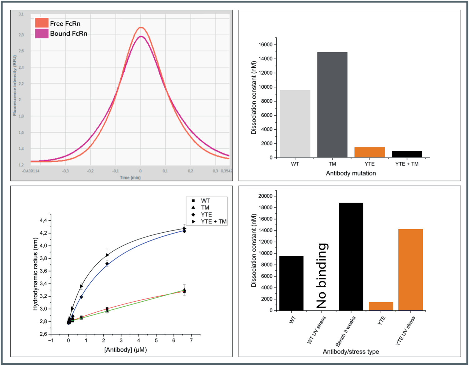
Top left: Taylor grams of free and bound ALC480-FcRn.
Top right: Affinities of mutated Fc against FcRn.
Bottom left: Binding curves of WT and mutant Fc.
Bottom right: Affinities of stressed Fc antibodies against FcRn
The WT and YTE Fc were subjected to UV radiation stress for FIDA. The WT Fc completely loses its ability to bind FcRn, while the YTE retains the ability, though the affinity is weakened 9.7-fold, demonstrating that the YTE mutation has increased the Fc’s ability to withstand UV stress.
The WT was also left at room temperature (RT) for 3 weeks, resulting in a 2-fold weakening of the affinity (Figure 2). Interestingly, when the WT Fc is subjected to UV radiation it exhibits aggregation characteristics that it did not have without the treatment, indicating that the UV treatment causes partial unfolding of the antibody or surface modifications of the protein. This effect is not present when the YTE mutant Fc is UV treated, indicating not only increased stability in binding functionality but also in resisting structural modifications (Figure 3, middle).
This demonstrates some of the many additional QC parameters of high relevance to drug development that are automatically identified during the FIDA assays.
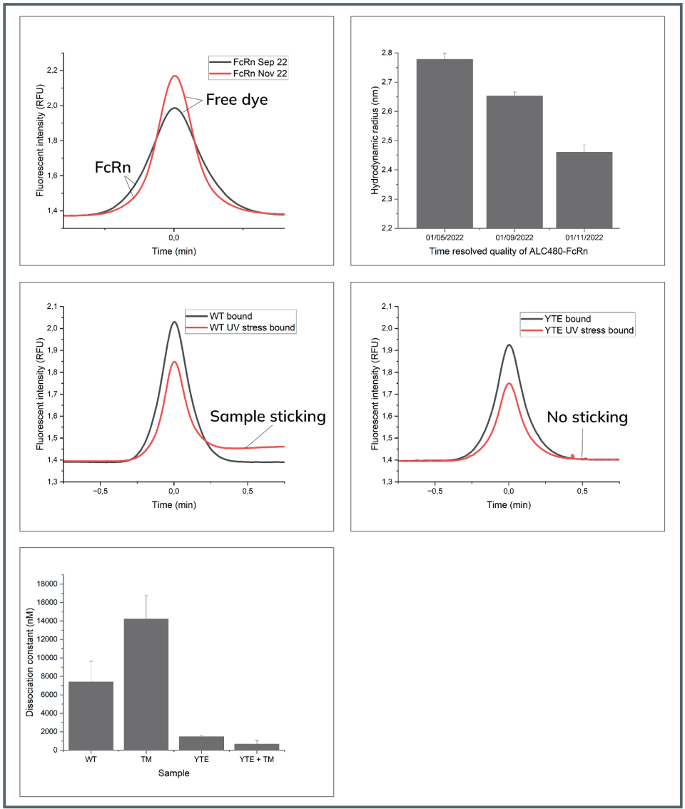
Top left: example Taylor grams of the same FcRn sample stored in the fridge.
Top right: Hydrodynamic radius of FcRn Stored at +4-8oC.
Middle left: Overlayed Taylor grams of WT Fc and UV stressed WT Fc antibody.
Middle right: Overlayed Taylor grams of bound YTE Fc and UV stressed bound YTE Fc antibody.
Bottom: Impact on reagent quality on the fitted Kds. The error bars represent the deviation between the Kd obtained in over the course of 7 months.
In the drug development process, quality control properties over the reagents is a necessary activity during CMC development.
To assess FIDA’s quality control properties over the reagents used in the experimental set-up, the ALC480-FcRn was stored for 6 months at 4-8oC, and the affinities against the mutant Fc’s were assessed.
The 4-8 C storage causes the ALC480-FcRn to lose its fluorescence label due to hydrolysis. This is not an issue in FIDA analysis but can cause issues with more conventional techniques. There is also a significant decrease in the size of the FcRn over time, indicating protein degradation (Figure 3, top right)
Encouragingly, the overall trend in the impact of the Fc mutations remains constant, demonstrating the power of Fida 1 to work even with suboptimal reagents (Figure 3, bottom).
CONCLUSION
FIDA enables in-solution FcRn assessment of the impact of Fc-specific sequence design and stress on antibody integrity and functionality during drug development. Critical changes to the structural properties of the antibodies can rapidly be identified while measuring affinities due to the simultaneous recording of several sample QC parameters. Demonstrating the broad applicability of FIDA during CMC development of antibodies.
REFERENCES
1 Properties of Human IgG1s Engineered for Enhanced Binding to the Neonatal Fc Receptor (FcRn). William F Dall’Acqua, Peter A Kiener, Herren Wu. J Biol Chem. 2006.
2 Structural characterization of a human Fc fragment engineered for lack of effector functions. Vaheh Oganesyan, Changshou Gao, Lena Shirinian, Herren Wu, William F Dall’Acqua. Acta Chrystallogr D Biol Crystallogr. 2008.