
Assessment of protein Stability and Functionality by Flow Induced Dispersion Analysis
INTRODUCTION
The present application note is based on a peer reviewed paper of 2019 (1). Protein-based pharmaceuticals represent a rapidly growing class of drug compounds. However, development of protein-based drugs is associated with significant challenges as these complex molecules are structurally labile. Further, the drug molecule, vehicle or degradation products may cause immunogenic responses, thereby leading to loss of therapeutic effect, toxicity or even anaphylaxis. Current methodologies cannot address these risks, as they typically are unable to probe stability under native conditions and require large amounts of sample.
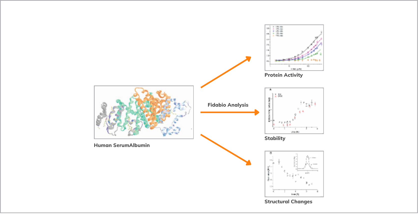
In this work, it is shown that Flow Induced Dispersion Analysis (FIDA) is used to measure the unfolding of Human Serum Albumin (HSA) and the loss of binding affinity to Fluorescein. The assay is performed under native conditions with minimal sample consumption. FIDA is a new capillary-based technology for measuring binding affinity and assessing protein stability in-solution under native conditions. FIDA utilises Taylor dispersion for accurate size determinations of analytes in a pressure driven flow system. The change in apparent size forms the basis for an accurate measure of binding affinity and protein stability.
Material & Methods
Fida 1 instrument with 266 nm and 480 nm LIF and LED fluorescence detection for unfolding and binding experiments respectively (Fidabio). FIDA standard capillary (i.d.: 75 μm, LT: 100 cm, Leff: 90 cm). 67 mM phosphate buffer pH 7.4 was used as working buffer. HSA (0.5 mg/mL) as indicator in 0-7 M urea for unfolding experiment.
Fluorescein as indicator (10 nM), HSA as analyte (0-500 μM) in 0-4 M urea solutions for binding experiments. Sample analysis was performed by filling the capillary with analyte, followed by injection of 39 nL indicator, which was mobilised towards the detector with analyte at 400 mbar.
Results
Urea-induced unfolding of HSA As FIDA technology provides absolute measurements of hydrodynamic radius, it was used to address global changes upon HSA unfolding. Additionally, the intrinsic fluorescence of HSA and its binding to a low molecular weight fluorescent ligand, Fluorescein, were probed to detect local structural changes and associated functionality of the protein in increasing urea concentration.
The changes in the size (hydrodynamic radius) of HSA was plotted as a function of increasing urea concentration (0-7 M) at 25 ̊C as shown in Figure 2A.
An increase in urea concentration around 4.0 M led to unfolding of HSA, observed as increase in size from 3.5 nm to 6.2 nm. The results are in line with a similar study using Small Angle X-ray Scattering (SAXS). The peak areas of FIDA taylorgrams were exploited for simultaneously probing the in-trinsic fluorescence intensity of HSA at in- creasing urea concentration.
Intrinsic fluorescence of HSA was affected by urea at 1.5 M indicating local structural changes prior to the overall unfolding (Figure 2B). This showcases the unique ability of FIDA to measure both local and global structural changes in a single measurement.
%20Unfolding%20curve%20of%20HSA....png)
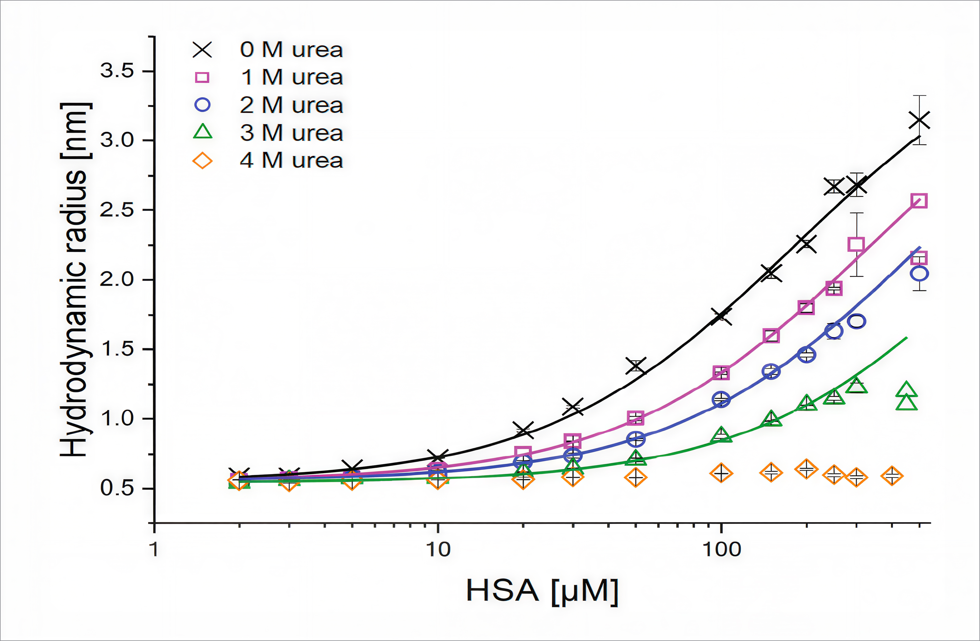
Loss of HSA binding to Fluorescein Binding curves of the interaction between Fluorescein and HSA in 0-4 M urea were generated (Figure 3) and the KD values were obtained as shown in Table 1. The binding affinity of the complex declines with increasing urea concentration; therefore, increased concentration of urea is associated with HSA unfolding and loss of functionality
.png)
Conclusions
A combination of different techniques is required for a complete understanding of protein stability and function. The FIDA methodology is a multitiered approach capable of detailed characterization of the denaturation and unfolding process as depicted by HSA unfolding induced by urea.
With minimal sample consumption and us- ing one instrument, FIDA allows in-depth assessment of protein activity combined with local and global structural changes by measuring the overall hydrodynamic radius of the protein, upon protein unfolding.
References
1. Morten, E. P.; Sarah, I. G.; Jesper, Ø.; Henrik, J., Protein characterization in 3D: size, folding and functional ASSSESSMENT in a unified approach. Anal. CHEM. 2019, 91, 4975-4979.
2. Jensen, H.; Østergaard, J., Flow Induced Dispersion Analysis Quantifies Noncovalent Interactions in Nanoliter SAMPLES. J. AM. CHEM. Soc. 2010, 132, 4070-4071.
3. Poulsen, N. N.; Andersen, N. Z.; Østergaard, J.; Zhuang, G.; Petersen, N.; Jensen, H., Flow Induced Dispersion Analysis Rapidly Quantifies Proteins in HUMAN PLASMA SAMPLES. Analyst 2015, 140, 4365-4369.
4. Poulsen, N. N.; Pedersen, M. E.; Ostergaard, J.; Petersen, N. J.; Nielsen, C. T.; Heegaard, N. H. H.; Jensen, H., Flow-Induced Dispersion Analysis for Probing Anti-dsDNA Antibody Binding Heterogeneity in Systemic Lupus Erythematosus Patients: Toward a New Approach for Diagnosis and Patient Stratification. Anal. CHEM. 2016, 88 (18), 9056-9061.
5. Abou-Zied, O. K.; SULAIMAN, S. A. J., Site-specific recognition of fluorescein by HUMAN SERUM ALBUMIN: A steady-state and TIME-resolved spectroscopic study. Dyes PIGMENTS 2014, 110, 89-96.