
Characterization of Conformational Changes by Flow Induced Dispersion Analysis
Introduction
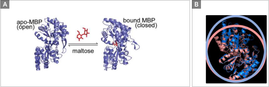
Many biological processes are regulated through the interactions of proteins with small molecules or other proteins. In many cases, these interactions induce conformational changes that directly modulate activities or provide new binding sites that facilitate building higher-order complexes. As a model system, we used Maltose Binding Protein (MBP), a member of the bacterial periplasmic binding protein superfamily.
MBP is the soluble component of the maltodextrin transport system and resides in the periplasm of Gram-negative bacteria, where it shuttles its ligands – maltose, maltotriose and maltoheptaose – to the membrane-bound transporter complex. The ligand-binding site of MBP is positioned between two domains separated by a hinge region.
Material & Methods
Fida 1 instrument with 480 nm LED fluorescence detection for binding experiments respectively (Fidabio ApS). FIDA standard capillary (i.d.: 75 µm, LT: 100 cm, Leff: 84 cm). Tris buffer pH 7.4 (20mM Tris, 150mM sodium chloride, 0.05% Tween) was used as the working buffer. MBP was used as indicator (4.3ugmL-1 , 100nM). MBP was labelled with an Atto 488 NHS ester from Sigma Aldrich. Maltose (O-α-D-Glucopyranosyl-D-glucose from Sigma M9171) was used as the analyte (0-1000 µM). Sample analysis was performed by filling the capillary with the analyte, followed by an injection of preincubated indicator and analyte, which was mobilized towards the detector with analyte at 400 mbar.
Results
Maltose induces a conformational change on the Maltose Binding Protein.
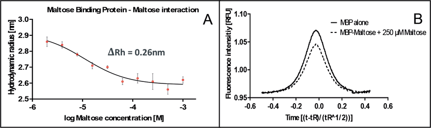
The FIDA technology provides an absolute measurement of hydrodynamic radius (Rh), and it was used to measure size changes of Atto488-labeled MBP (42.5 kDa) upon structural change srelated to binding to Maltose (0.3 kDa). The change in apparent Rh of MBP was plotted as a function of increasing Maltose concentration (0-1000 µM) at 25°C as shown in Figure 2A. The Rh of MBP decreases from 2.88nm to 2.62nm which corresponds to a ΔRh of 0.26nm, clearly indicating a structural change upon binding (Figure 2A). The evaluated Kd for this interaction is in the range of 10 µM which corresponds to the literature [1,2]. In Figure 2, the overlay FIDA signal of MBP and MBP-Maltose is shown. In Figure 2B, the indicator peak gets narrower in the presence of Maltose. The peak areas of FIDA taylorgrams were exploited for simultaneously probing the fluorescence intensity of MBP at increasing Maltose concentration. It shows that the fluorescence of MBP was affected by Maltose (Figure 2B), allowing an orthogonal estimate of binding (data not shown).
Conclusion
The presented data show how conformational changes of proteins can be measured in-solution using the FIDA technology. FIDA provides in-depth assessment of activity combined with local and global protein structural changes by measuring the overall hydrodynamic radius of the protein with minimal sample consumption (5 µL). In addition, it is possible to measure the affinity constant for the analyzed interaction.
References
1. Shahir S Rizk et al.; Allosteric control of ligand-binding affinity using engineered conformation-specific effector proteins., 2011, Natural structure & molecular biology; Vol 18 No. 4, 2011.p. 437-444.
2. Shahir S. Rizk et al.; Allosteric Control of Ligand Binding Affinity Using Engineered Conformation-Specific Effector Proteins., 2011 April; 18(4): 437–442. doi:10.1038/nsmb.2002.