
Detection of Auto-Antibodies in Systemic Lupus Erythematosus Patients
Introduction
Systemic Lupus Erythematosus (SLE) is an autoimmune disease, where the detection of circulating auto-antibodies against ds-DNA is used as an important diagnosis criterion. However, current methods for detecting anti-ds-DNA antibodies in blood samples are primarily based on ELISA procedures utilizing surface chemistries (i.e. immobilization of antigen), multiple steps and cumbersome optimization. Here we present a simple and immobilization-free procedure, termed Flow-Induced Dispersion Analysis (FIDA), for detecting auto-antibodies against ds-DNA directly in 85 % plasma samples.
FIDA is a new capillary-based technology for measuring in-solution binding under native conditions in complex solutions (e.g. plasma). FIDA is based on Taylor dispersion in a pressure driven flow of a ligand (termed indicator, e.g. ds-DNA) interacting with the analyte of interest (e.g. anti-ds-DNA anti-body). The indicator alone appears small (i.e. it has a small hydrodynamic radius) when it is not bound to the antibody, but upon binding it will appear larger (i.e. the complex has a larger hydrodynamic radius). The change in apparent size forms the basis for an accurate measure of analyte concentration and interaction.
Material & Methods
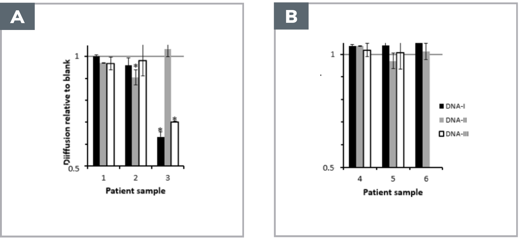
488 nm Laser-Induced Fluorescence detection. FIDA-coated capillary (i.d.: 75 μm, LT: 75 cm, Leff: 65 cm). 100 mM phosphate buffer pH 7.9 was used as working buffer, three different ds-DNA sequences (32 bp) labelled with atto488 was used as indicators (50-100 nM), monoclonal antibody against ds-DNA (0-1000 nM) was spiked into 0-85 % human plasma (healthy control) and used as analyte, 85 % plasma from six SLE patients was used as analyte [1].
Sample analysis was performed by filling the capillary with an analyte-zone, followed by injection of 26 nL indicator, which was mobilized towards the detector with analyte at 50 mbar.
Results
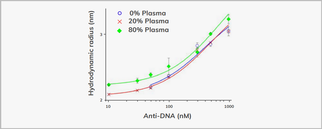
The apparent size (hydrodynamic radius) of the ds-DNA indicator was plotted as a function of antibody concentration (Figure 1, 0-1000 nM) in 0, 20 and 85 % healthy donor plasma respectively. The obtained dissociation constants (Kd) were 236 nM, 278 nM and 362 nM in 0, 20 and 85 % plasma respectively, and thus correlated well.
References
1. Poulsen NN, Pedersen ME, Østergaard J, et al (2016) Flow-Induced Dispersion Analysis for Probing Anti-dsDNA Antibody Binding Heterogeneity in Systemic Lupus Erythematosus Patients: Toward