
Easy and Rapid Assessment of Protein ubiquitination, e.g. in PROTAC development
Introduction
Ubiquitination is one of the common and most important posttranslational modifications in eukaryotic cells. Ubiquitination functions as a regulatory mechanism that controls processes ranging from protein stability, protein degradation to membrane transport and transcription. The process is carried out by recruitment of three key enzymes: ubiquitin activating enzyme E1, ubiquitin-conjugating enzyme (E2) and ubiquitin ligase (E3) (1,2). Many variants of these enzymes exist in nature and have specificity towards their target protein
This application note presents a quick assay to determine protein ubiquitination using Fida 1. Fida 1 is a capillary-based technology for measuring in-solution binding and complex size of biomolecules.
In the present work, a model system based on P53 ubiquitination was used to develop the assay. As compared to conventional and commercial ubiquitination protocols that require running of SDS-PAGE and western blots, Fida 1 provided clear results much faster and using small volumes.
Material & Methods
Fida 1 instrument with 480 nm LED fluorescence detection was used for assay development (Fida Biosystems ApS) with Fidabio standard capillary (i.d.: 75 μm, LT: 100 cm, Leff: 84 cm). HEPES buffer, pH 7.5, 500mM NaCl, 10mM TCEP was used as the working buffer. P53, E1, E2, E3 and ATP were mixed in vials in a final concentration of 100nM, 50nM, 1uM, 1uM and 10mM, respectively.
Ubiquitin was added in varying Ub) Ubiquitin concentrations to manipulate the degree of ubiquitination. 1 uM fluorescently labelled (fl-was used in the assay as a tracer. The reactions were incubated for 15 mins and 1 hour. Dispersion-Analysis was performed by injecting the capillary with the analyte (buffer) (4μL), followed by an injection of 39 nL of reactions mix with fl-Ub as indicator and mobilised towards the detector at 400 mbar.
Results
Size determination of Fl.Ubiquitin
The FIDA technology provides absolute measurement of molecular size (hydrodynamic radius) for a labelled molecule called indicator. In this assay fl-Ub serves as an indicator. The hydrodynamic radius of fl-Ub was determined to be 1.8 nm.
Assay Setup
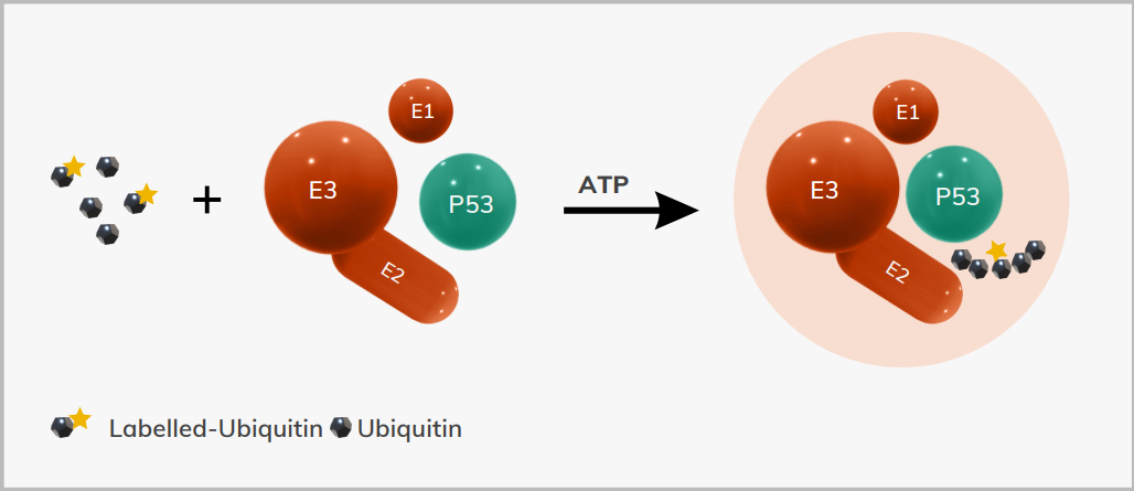
1 uM of fl-Ub was added to the ubiquitination reaction as a tracer molecule. The reaction is designed for ubiquitination of P53 protein and all the relevant enzymes (E1, E2, E3), ATP, fl-Ub and unlabelled (free) ubiquitin was added to it. An increase in the size of fl-Ub confirms the recruitment into ubiquitination of P53 (figure 1).
The ubiquitination reaction of P53 was carried out with varying concentrations of unlabelled ubiquitin to assess and manipulate the degree of ubiquitination. The reactions were incubated at 37 ̊C for 15 mins and one hour.
A negative control experiment where no ATP was added was performed to ensure no ubiquitination reaction in the given conditions.
The results showed that there was no change in the size of the fl-Ub in the negative control, however in the positive reactions the size of the fl-Ub changed from 1.8nm to 16 nm and 21nm, where 20uM and 50uM free ubiquitin was added to the reactions respectively (figure 2, Table 1).
The change in the size was eminent after 15 mins of incubation. As it is known that complete ubiquitination takes place in 10-15 minutes at 37 ̊C, no increase in the complex size was observed after one hour of incubation (Table 1 on the next page).
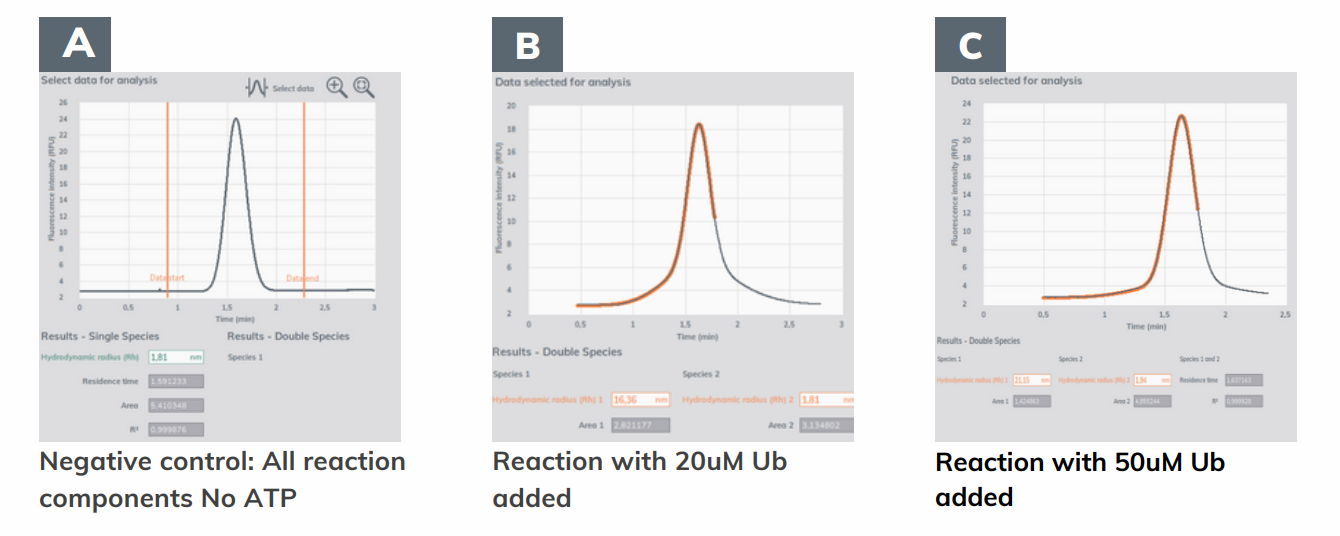
Figure 2.
A) Negative control for ubiquitination reaction which has all components added except ATP.
B) Ubiquitination reaction with all components and 20uM of free ubiquitin (Ub) added.
C) Ubiquitination reaction with all components and 50uM of free ubiquitin (Ub) added.
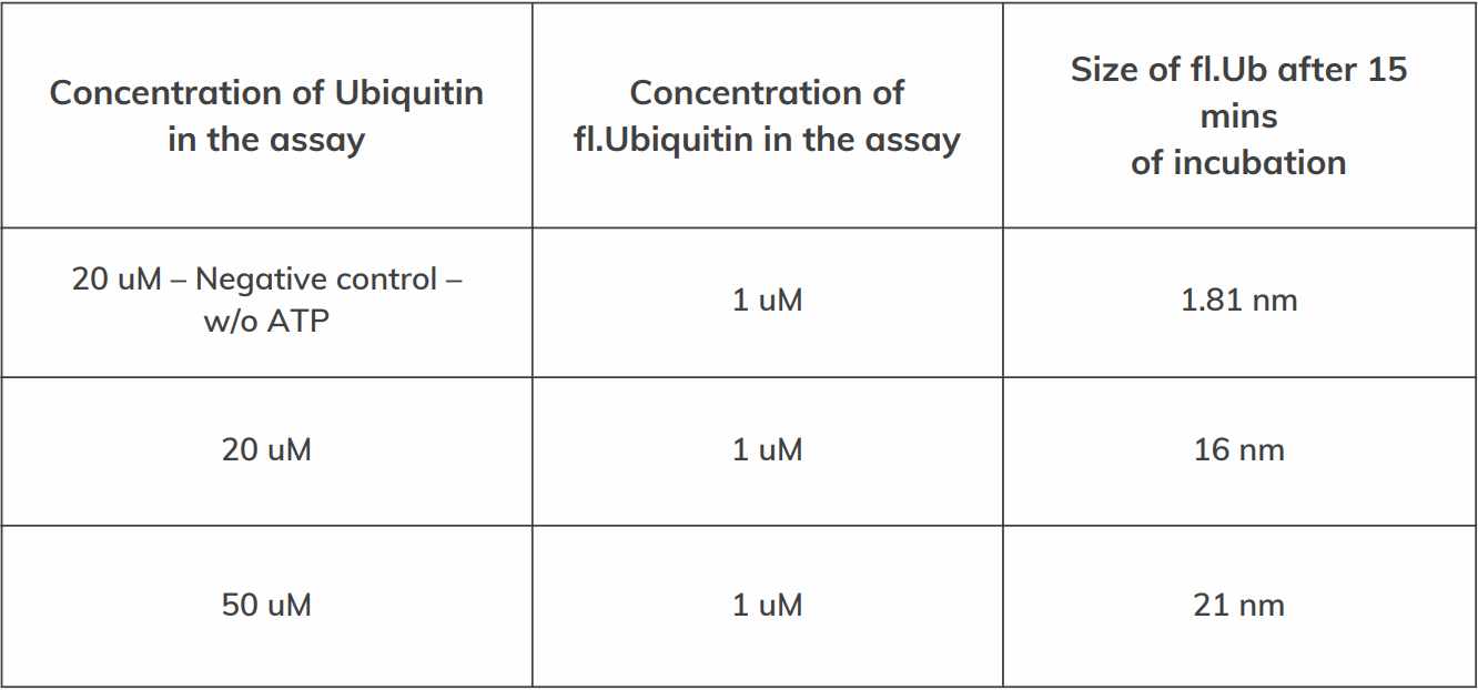
Table 1: Increase in the size of fl.Ub upon recruitment into ubiquitination of P53. The size of the fl.Ub alone was measured to be 1.81nm. It can be seen that the size of the fl.Ub did not increase in a negative control reaction (with no ATP) added implying that any increase in the size of fl.Ub occurred only when the ubiquitination of P53 took place.
Conclusions
The FIDA technology enables quick and easy assessment of ubiquitination reaction for the protein of interest. As compared to the commercial kits, where SDS-PAGE and western blots are required to assess ubiquitination, FIDA provides an opportunity for real-time monitoring of the ubiquitination reaction as well as quick running times ranging from 15-30 mins. Thus, substantially reducing the assay times.
References
1. Zhao Q.; Liu L.; Xie Q.; Chapter 13, In Vitro Protein Ubiquitination Assay. Springer Protocols. 2012
2. Vikki M. Weake; Jery L.Workman; Handbook of cell signaling, Ubiquitination. 2010