From fibril to monomer: understanding amyloid stability with FIDA
Thermodynamic stability of amyloid fibrils
Amyloid fibrils accumulate in neurodegenerative diseases including Alzheimer's and Parkinson's, which has prompted extensive study of the kinetics and mechanisms of their formation. Yet knowing how fibrils form, or what they look like, doesn't fully explain why different fibrillar structures emerge, how they respond to their environment, or how readily they can be disrupted.
The thermodynamic stability of the resulting structures has received comparatively little attention, despite providing valuable insight into the energetic differences between amyloid polymorphs and the conditions that favour them.1 These differences in stability are consequential to the effectiveness of biological clearance mechanisms, with a recent study reporting that more stable fibrils were more resistant to disaggregation by molecular chaperones.2
Understanding what makes existing fibrils stable, and how that stability might be altered, provides a route to therapeutic strategies aimed at promoting fibril disassembly and reversing amyloid accumulation.
FIDA offers a practical solution for investigating this property, directly quantifying soluble protein in fibril-containing mixtures following chemical depolymerisation, without requiring fluorescent labels or preparative separation.
The challenge: Balancing confidence and convenience
Measuring amyloid fibril stability is not as straightforward as monitoring fibril formation. Thermodynamic stability is determined by the concentration of soluble protein in equilibrium with fibrils, which can be exceptionally low for highly stable aggregates. Chemical depolymerisation can shift this equilibrium to make the soluble fraction measurable, but a fundamental analytical challenge remains: how can soluble protein be accurately quantified in a heterogeneous mixture containing fibrils?
Conventional methods involve compromises between direct quantification, practical efficiency and applicability. Separation-based techniques such as ultracentrifugation enable soluble protein to be quantified, but can be laborious, time-consuming and sample-intensive. Faster, more scalable alternatives, including Thioflavin T fluorescence, light scattering and differential scanning fluorimetry, rely on indirect signals that can be sensitive to fibril structure, solution conditions or protein composition. These dependencies introduce uncertainty and limit applicability: a convenient measurement for one amyloid system may yield ambiguous results for another.1
Confidence in the measurement matters just as much as convenience. The challenge, therefore, is to combine dependable quantification of soluble protein with an efficient analytical workflow, without relying on extensive sample preparation or indirect measurements that may not be suitable for every fibril preparation.
The approach: Direct quantification through transient incomplete separation
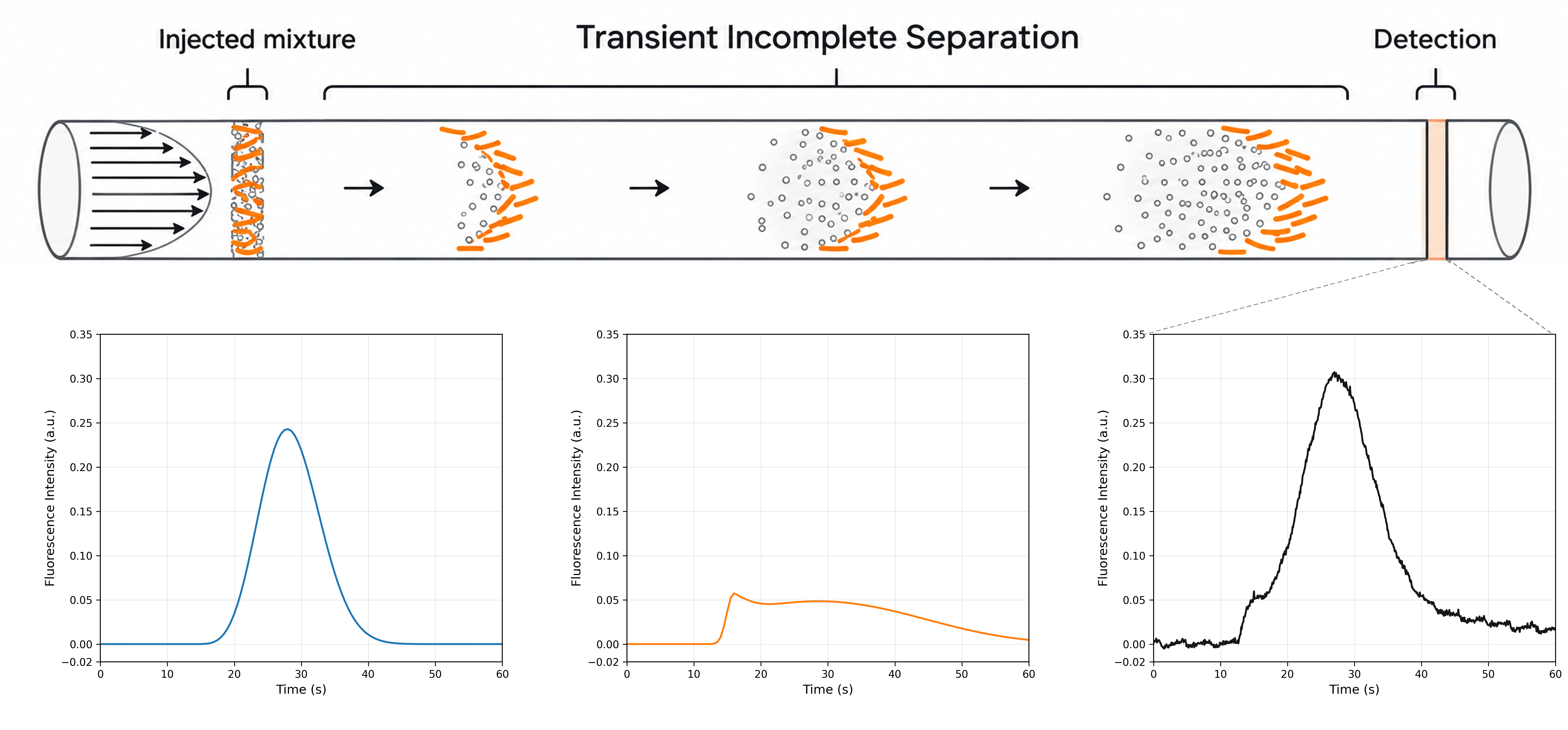
FIDA addresses this challenge by directly quantifying the soluble protein fraction in a fibril-containing mixture. The method uses transient incomplete separation (TIS), in which soluble monomers and amyloid fibrils behave differently as they disperse through a capillary under laminar flow. This produces distinct signal profiles, allowing the soluble protein population to be measured without preparative separation of fibrils from monomer. In the reported workflow, soluble protein was detected label-free using its intrinsic fluorescence.
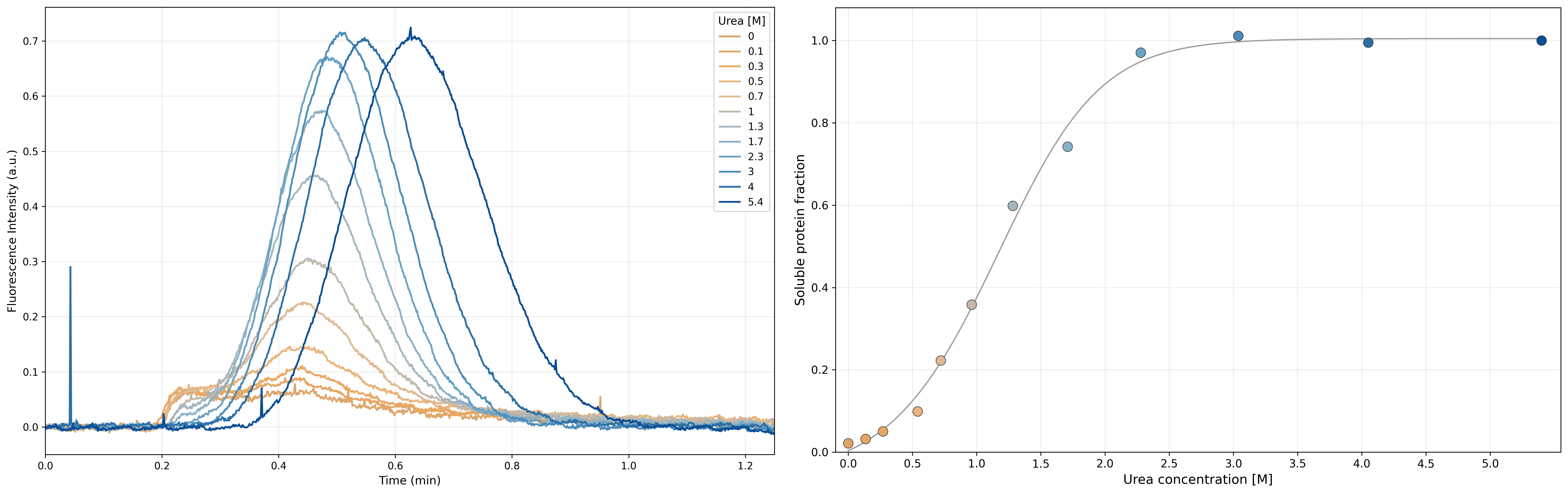
To determine fibril stability, this measurement is combined with chemical depolymerisation. Fibrils are incubated across a denaturant series, shifting the equilibrium between soluble protein and fibrillar material. FIDA then quantifies the soluble fraction at each condition, generating a depolymerisation curve from which the thermodynamic stability of the fibrils can be derived. Rather than relying on a fibril-associated optical signal, the approach directly tracks the soluble protein population that defines the equilibrium.
The result: Reliable stability measurements across amyloid systems
Farzadfard and colleagues evaluated the FIDA approach against established techniques using three amyloid systems: wild-type α-synuclein, its F94W variant and PI3K-SH3. FIDA successfully generated quantitative depolymerisation profiles for all three, with stability measurements agreeing closely with established methods where applicable.1
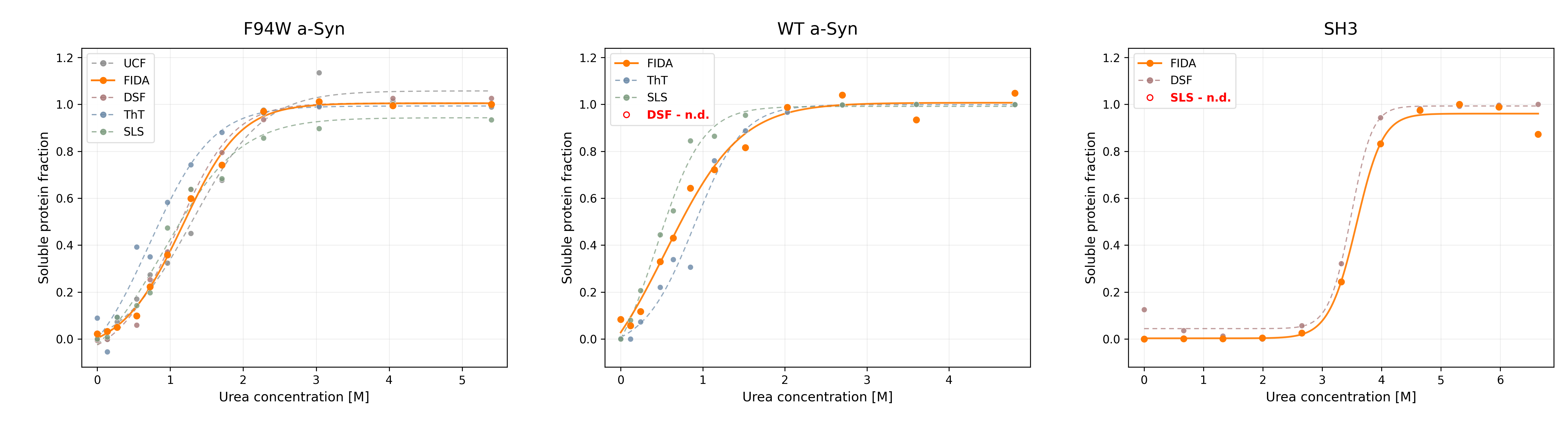
For F94W α-synuclein, FIDA measured a Gibbs free energy (ΔG) of −34.6 ± 1.0 kJ/mol, closely matching ultracentrifugation (−34.0 ± 0.9 kJ/mol) and differential scanning fluorimetry (−35.4 ± 1.2 kJ/mol).
Importantly, FIDA also quantified the stability of wild-type α-synuclein without introducing a fluorescent probe or modifying the protein, where differential scanning fluorimetry could not provide a meaningful measurement. Similarly, FIDA produced stability measurements for PI3K-SH3 that agreed with differential scanning fluorimetry, while light scattering proved unreliable.
Together, these comparisons demonstrate that direct, label-free quantification of soluble protein enables FIDA to deliver stability measurements across different amyloid systems, including where conventional methods encounter limitations. The workflow combines this analytical capability with practical efficiency: approximately 15 automated measurements per hour, less than 250 nL consumed per measurement, and sensitivity sufficient to quantify wild-type α-synuclein monomer down to approximately 5 µM using intrinsic tyrosine fluorescence.1 Crucially, the convenience of the workflow does not come at the expense of confidence in the measurement.
The insight: From amyloid polymorphism to disaggregation
The fibrils that form fastest are not necessarily the most stable, and greater stability may make existing fibrils harder to disassemble. By incorporating FIDA-based stability measurements into their research, scientists are uncovering the energetic differences behind amyloid structure, formation and disassembly.
Using FIDA, Farzadfard and colleagues found that α-synuclein fibrils assembled with and without salt exhibited similar stability under their respective preparation conditions. Yet when measured in the same salt-containing environment, the polymorph formed without salt was approximately 8.5 kJ/mol more stable, despite its slower formation kinetics. FIDA also revealed that fibrils assembled under acidic conditions became less stable when transferred to neutral pH. These findings demonstrate how fibril polymorphism and environmental conditions influence stability in ways that aggregation kinetics alone cannot explain.1
These differences have biological consequences. Fricke and colleagues used FIDA to investigate whether stability influences susceptibility to disaggregation, and found maturation of one α-synuclein fibril preparation increased its stability by approximately 10 kJ/mol while reducing its susceptibility to molecular-chaperone-mediated disaggregation by around 50%.2
The applications are expanding beyond these initial investigations: FIDA-derived stability has informed how mutations alter polymorphism,3 and been incorporated into characterisation of underexplored neurodegenerative proteinopathies.4 Together, these studies show how measuring thermodynamic stability can deepen our understanding of amyloid behaviour and help identify the molecular and environmental factors that could inform the development of therapeutic interventions aimed at improving amyloid clearance.
Conclusion: Making fibril stability more accessible
Once amyloid fibrils have formed, understanding what keeps them intact is essential to investigating how they might be disrupted. Thermodynamic stability offers a quantitative measure of the energetic differences between fibrillar structures, helping researchers understand their persistence, response to environmental changes and susceptibility to disassembly. This makes stability a valuable parameter not only for characterising amyloid accumulation, but also for investigating how it might be reversed.
By bringing direct, label-free quantification of soluble protein into an efficient, automated workflow, FIDA makes this dimension of amyloid characterisation more accessible. Researchers can now investigate the factors governing fibril stability without having to compromise between practical efficiency and confidence in their measurements, generating insights that could inform new strategies for destabilising pathological amyloids.
References
- Farzadfard A, Kunka A, Mason TO, et al. Thermodynamic characterization of amyloid polymorphism by microfluidic transient incomplete separation. Chemical Science. 2024;15:2528–2544. https://doi.org/10.1039/D3SC05371G
- Fricke C, Kunka A, Norrild RK, et al. Thermodynamic stability modulates chaperone-mediated disaggregation of α-synuclein fibrils. Chemical Science. 2026;17:283–298. https://doi.org/10.1039/D5SC04927J
- Kunka A, Farzadfard A, Larsen JA, et al. Probing the energy landscape of α-synuclein amyloid fibril formation by systematic K-to-Q mutagenesis. Advanced Science. 2026;e77643. https://doi.org/10.1002/advs.77643
- Konstantoulea K, Gadhe L, Goodavish F, et al. TAF15 amyloids propagate via defined motifs in a prion-like fashion. Nature Communications. 2026;17:8625. https://doi.org/10.1038/s41467-026-75607-3
VERSION 1.0