From monomer to fibril: understanding amyloid growth with FIDA
The formation and growth of amyloid fibrils are central to the study of protein aggregation in neurodegenerative diseases such as Alzheimer's disease, making them important targets of therapeutic research. A 2026 review of the Alzheimer's drug development pipeline found that approximately 25% of investigational drugs targeted intervention in amyloid-β or tau-related processes.1
Understanding these processes requires insight into the physical development of fibrillar assemblies. How quickly do fibrils grow? When does their physical growth slow or stop? How does that change in response to experimental conditions or therapeutic interventions?
FIDA offers a different perspective on these questions: the ability to measure hydrodynamic size in solution, revealing how the physical dimensions of molecular assemblies change.
The challenge
The aggregation signal has plateaued, but have your fibrils?
Thioflavin T (ThT) fluorescence is widely used to monitor aggregation kinetics, but fluorescence intensity is not a direct measurement of fibril dimensions. Transmission electron microscopy (TEM) provides detailed images of fibril length and morphology, but its limited scalability and challenges in identifying very short fibrils constrain repeated quantitative analysis. Both provide important perspectives, but what remains difficult is following changes in fibril-associated size quantitatively, in solution, across the aggregation time course.
FIDA adds that missing dimension: an in-solution, size-sensitive measurement of fibril assemblies. In a published Tau repeat-domain (TauRD) study,2 FIDA revealed continued growth after the corresponding ThT trace had plateaued, progressing the analytical question from "Has amyloid formed?" to "What is happening to the fibrillar assemblies in solution?"
The approach
Following fibril growth in solution
FibrilPaint is a family of fluorescently labelled peptides developed by Aragonès Pedrola, Dekker and colleagues to recognise amyloid fibrils.3 FibrilPaint1 exhibits high-affinity binding to fibrillar Tau without detectable binding to monomeric TauRD in the reported experiments,2 while subsequent research has demonstrated fibril recognition across additional peptide variants and amyloid systems.
Combined with FIDA, FibrilPaint provides a way to investigate how fibrillar assemblies develop in solution. When the fluorescent peptide associates with an amyloid fibril, its diffusion behaviour reflects that of the larger assembly. FIDA translates this into a measurement of hydrodynamic radius (Rh), providing a quantitative, size-sensitive readout of fibril-associated material. By sampling an aggregation reaction at successive timepoints, researchers can follow how this physical dimension changes as fibrils develop.
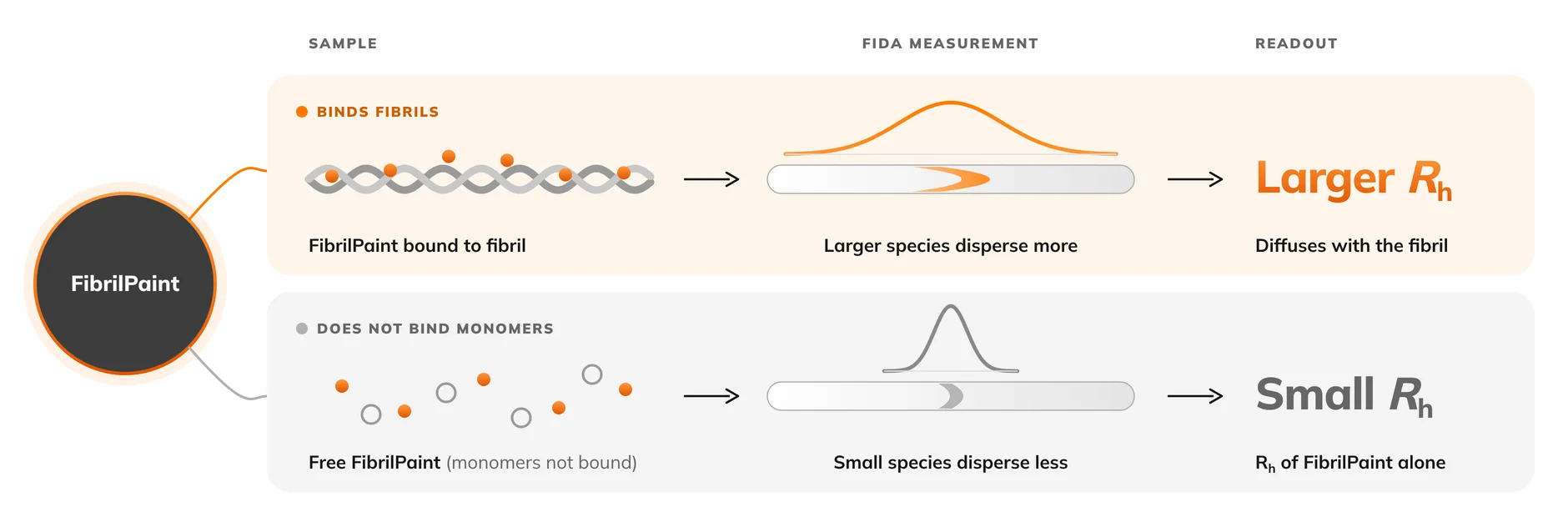
Figure 1. Measuring fibril-associated size using FibrilPaint and FIDA. When fluorescent FibrilPaint binds to an amyloid fibril, it diffuses with the larger complex, resulting in an increased measured hydrodynamic radius (Rh; orange). Unbound FibrilPaint, which does not bind monomers, retains its smaller hydrodynamic radius (grey).
Image credit: Adapted from Figure 1 of Aragonès Pedrola et al.2 © 2025 the authors. Licensed under CC BY 4.0. Original publication: https://doi.org/10.1073/pnas.2502847122.
To demonstrate the approach, Aragonès Pedrola and colleagues investigated heparin-induced aggregation of recombinant TauRD carrying the ΔK280. 20 µM TauRD was aggregated with 5 µM heparin at 37 °C, with FibrilPaint1 introduced to samples at selected timepoints for FIDA analysis. In parallel, ThT fluorescence and transmission electron microscopy provided complementary perspectives:
- ThT: the progression of amyloid-sensitive fluorescence.
- FIDA + FibrilPaint1: changes in fibril-associated hydrodynamic size in solution.
- TEM: direct visualisation of fibril morphology and length.
All three techniques examined the same aggregation process, but revealed different insights into fibril development.
The result
Growth beyond the fluorescence plateau
The ThT signal increased rapidly, approaching a plateau after approximately 2 hours and showing little further change beyond 4 hours. By contrast, the hydrodynamic radius of TauRD fibril-associated species measured by FIDA continued to rise, from approximately 10 nm beyond the 4-hour mark to 45 nm at around 8 hours (Fig. 2A–B).2
The FIDA size trajectory therefore revealed a stage of continued fibril-associated growth that was not apparent from the ThT fluorescence trace alone.
TEM provided independent morphological evidence supporting continued fibril elongation. The authors reported mean fibril lengths increasing from approximately 140 nm after 2 hours, to 290 nm after 5 hours and 580 nm after 24 hours in their sampled TauRD preparations (Fig. 2C–D).
For very early structures, however, microscopy faced a practical limitation: the limited visible structure of fibrils shorter than approximately 80 nm prevents their reliable identification and inclusion in the TEM analysis. By contrast, FIDA detected an early hydrodynamic radius of approximately 4 nm after just 1 hour, corresponding to an estimated fibril length of just 2 nm (four fibril layers) using the authors' structural model.2
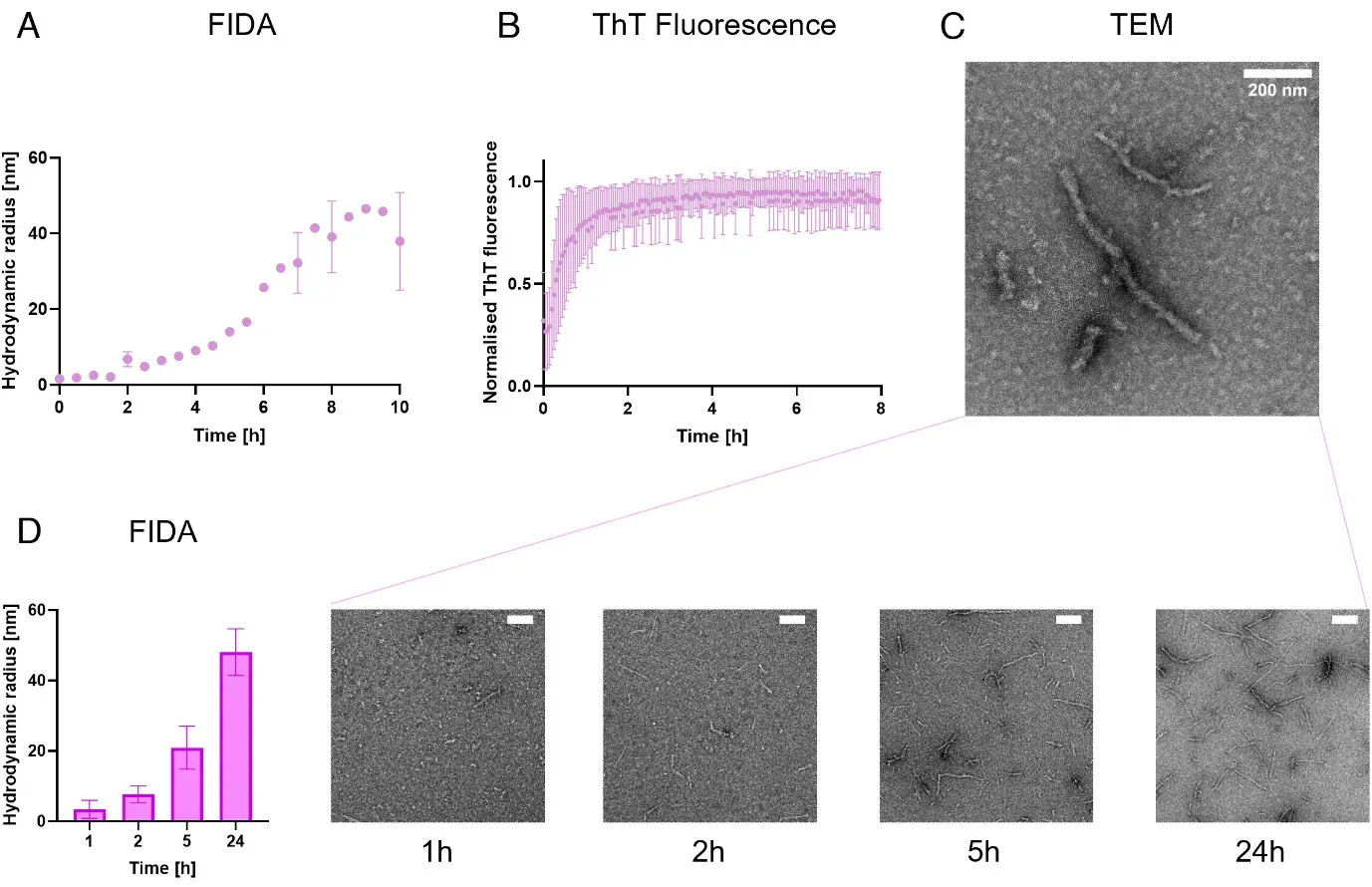
Figure 2. Following TauRD fibril growth with FIDA, ThT and TEM. (A) FIDA measurements of fibril-associated hydrodynamic radius during TauRD aggregation, showing continued size increases after (B) the ThT fluorescence signal approaches a plateau. (C) Representative TEM images of TauRD fibrils after 24 hours (scale bars: 200 nm). (D) Comparison of FIDA-measured hydrodynamic radius and TEM-measured fibril length at 1, 2, 5 and 24 hours. Panels A and B show representative FIDA data (N = 3) and averaged ThT triplicates (N = 3), respectively; panel D shows averaged duplicate measurements.
Image credit: Reproduced without modification from Figure 3 of Aragonès Pedrola et al.2 © 2025 the authors. Licensed under CC BY 4.0. Original publication: https://doi.org/10.1073/pnas.2502847122.
The insight
What fibril size reveals
In the TauRD experiments, TEM could not reliably identify fibrils shorter than 80 nm, while ThT fluorescence was already approaching its plateau when fibrils had reached a length of just 4.7 nm, as estimated from FIDA measurements.
FIDA detected even earlier TauRD assemblies corresponding to approximately four fibril layers (2 nm in length), while separate measurements of patient-derived Alzheimer's fibrils extended to an estimated 1,100 layers (510 nm in length).2 This breadth offers researchers a way to investigate fibril development from the earliest assemblies all the way through to mature fibrils, spanning stages that conventional aggregation fluorescence and microscopy cannot readily characterise together.
Following fibril-associated size throughout the entire aggregation allows researchers to identify both when and how conditions or interventions alter fibril development. The ability to observe whether a compound delays the onset of growth, slows its subsequent progression, or changes the size ultimately reached by the fibrils provides valuable insight into the underlying mechanism of action. An intervention affecting early fibril development raises different mechanistic questions from one that alters later growth, while a reduction in fibril-associated size could prompt investigation of fragmentation or disassembly.
By identifying where changes occur along the growth trajectory, FIDA goes beyond detecting an effect towards understanding how that effect arises, better informing the development of aggregation-modifying strategies.
Conclusion
Bringing fibril growth into focus
Investigating how conditions or potential therapeutics affect fibril growth requires measurements across multiple timepoints, samples, and conditions. FIDA's distinctive combination of automation, low sample consumption, and broad in-solution size range offers a practical route to performing aggregation studies at scale, from monomer to fibril.
The perspective gained from direct observation of size allows insight beyond just the onset of aggregation; it provides the opportunity to view how fibrils develop, how their growth can be influenced, and where interventions make a difference. For researchers seeking to understand or control amyloid aggregation, FIDA makes fibril size a quantitative, scalable part of the experimental workflow.
References
- Cummings, J. L., Zhou, Y., Yang, Y., et al. (2026). Alzheimer's disease drug development pipeline: 2026. Alzheimer's & Dementia: Translational Research & Clinical Interventions, 12(2), e70251. https://doi.org/10.1002/trc2.70251
- Aragonès Pedrola, J., Dekker, F. A., Garfagnini, T., et al. (2025). FibrilPaint to determine the length of Tau amyloids in fluids. Proceedings of the National Academy of Sciences, 122(44), e2502847122. https://doi.org/10.1073/pnas.2502847122
- Aragonès Pedrola, J., Dekker, F. A., Guttmann, K., et al. (2026). Fibrilpaint: A class of amyloid-targeting peptides. Chemistry – A European Journal, e71539 (early online publication). https://doi.org/10.1002/chem.71539