
Functional Characterization of Nanodisc embedded GPCR
Introduction
G protein-coupled receptors (GPCRs) are the largest family of membrane proteins, and they are involved in many vital physiological cell mechanisms, e.g., hormonal signalling, cell cycle regulation, neurotransmission signalling and odour receptors. GPCRs-targeting drugs represent 27% of the global commercial drugs. Additionally, GPCRs are particularly challenging to work with, given their instability and small-molecule ligands that are difficult to label. Therefore, there is an increasing demand for new technologies to advance discoveries and ultimately new treatments for GPCR related disorders.
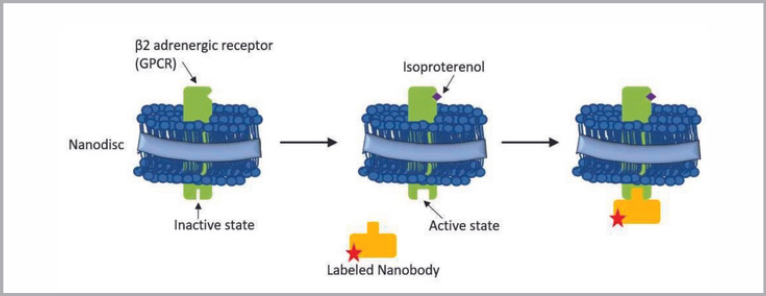
This work demonstrates that Flow-Induced Dispersion Analysis (FIDA) can be used to fully characterise the binding between Nanodisc-embedded β2-adrenergic receptor and Nanobody (Nb80). Further, Fida 1 has a built-in quality control making it possible to assess the receptor’s oligomeric state and the integrity of the sample.
FIDA is a new capillary-based technology for measuring binding affinity through absolute and accurate size determinations of analytes in a pressure-driven flow system. The change in apparent size forms the basis for the accurate analysis/determination of binding affinity and protein stability.
Material & Methods
FIDA 1 instrument with 480 nm LED fluorescence detection for binding experiments (Fidabio ApS). Fida standard capillary (i.d.: 75 μm, LT: 100 cm, Leff: 84 cm). HEPES buffer 10mM pH 7.4, 0.5 mg/ml BSA was used as the working buffer. Nanobody Nb80 was used as the indicator. Nb80 was labelled with an Alexa Fluor® 488 Protein Labelling Kit from ThermoFisher Scientific
NanoDisc-embedded β2-adrenergic receptor (β2AR) was used as the analyte (0-1 μM). Sample analysis was performed by filling the capillary with the analyte (4μL), followed by an injection of 39 nL of pre-incubated indicator+analyte, which was mobilised towards the detector at 400 mbar.
Results
Nb80 binds to the active GPCR.
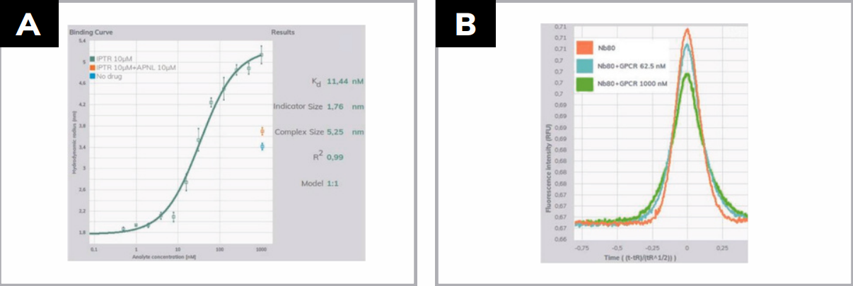
The FIDA technology provides an absolute measurement of the hydrodynamic radius (Rh), and in this work, it was used to measure the size change of Alexa488-labeled Nb80 upon binding to increasing concentration of Nanodiscs-embedded β2AR (ND-β2AR). The change in apparent Rh of Nb80 was plotted as a function of increasing NDβ2AR concentration (0-1 µM) in presence of isoproterenol 10 µM, an agonist that binds to the extracellular side of the GPCR and turns it into the active state (Figure 2A, green line). The Rh of Nb80 without ND-β2AR was 1,76 nm, and upon addition of ND-β2AR the size increased up to 5.25 nm, clearly indicating a binding event.
The Rh of 5.25 nm is in agreement with the average size of these Nanodiscs, which is around 5nm. To demonstrate the specificity of the binding, 2 additional controls were measured. The first one was in the presence of an antagonist, alprenolol (APNL) 10µM, which turns the receptor into the inactive state (Figure 2A, orange dot). The second control was a measure of labelled nanobody in the presence of ND-β2AR but without agonist nor antagonist (Figure 2A, blue dot). The two controls Rh at 3.6 and 3.4 strongly demonstrate the reduced binding ability of the GPCR when it is in the inactive state.
Conclusions
The data shows how FIDA can characterise Nanodisc-embedded proteins, in solution and non-invasively.
Besides receptor binding, FIDA technology also provides the possibility of assessing dimerisation and oligomerisation of GPCRs, and it can work with any scaffold up to 500 nm radius, such as proteoliposomes.