
High throughput detergent screening for membrane proteins
INTRODUCTION
More than 50% of the present-day drugs, and an increasing number of drug discovery and development programs target proteins situated in a membrane. Membrane proteins are challenging to express, maintain in-solution, purify, and stabilise after extraction from cells. As a result, most of the membrane proteins undergo detergent screening to identify the detergents that solubilise the highest amount of protein from the insoluble fraction of cell lysate. Effective detergents offer high degree of solubilisation as well as stabilisation of membrane proteins.
In this Application Note, Fida 1 is used for high throughput detergent screening to assess the degree of solubilisation and polydispersity of a GFP-tagged membrane protein directly in crude matrix, using only 40nL of sample per measurement. Moreover, the FIDA detergent screening is compared with Fluorescence Size- Exclusion Chromatography (FSEC)
FIDA MEASURING PRINCIPLE
Flow-Induced-Dispersion-Analysis (FIDA) is a capillary-based microfluidic method, exploiting that flow rates in the centre of the capillary are faster than at the edges of the capillary. The resulting radial concentration gradients at the front and the tail of the sample zone result in the diffusion of your chosen indicator. The distinct dispersion profiles determine the shape of the generated curves and enables a “first principle” biophysical measurement of size (Figure 1). The FIDA measurement of size is broadly applicable for studying biomolecular stability, interactions, etc.
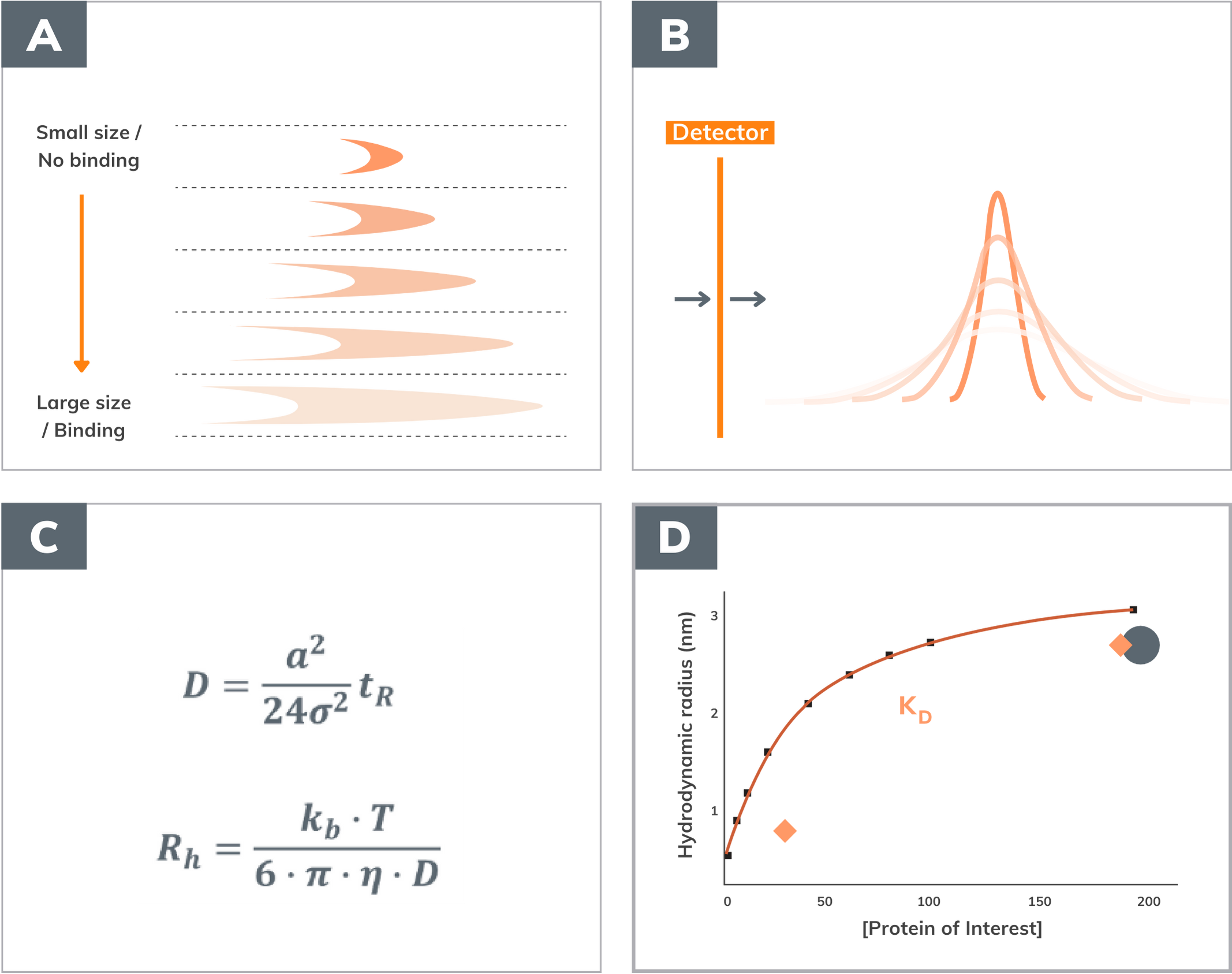
A. Dispersion profiles occurring in capillary. The shape of the profile depends on the size of the fluorescent molecule or on the size of the complexes it forms with its binding partners
B. Corresponding detector readout,
C- D. Calculation of diffusivity, D, and hydrodynamic radius, Rh, is used to measure the affinity, KD, of binding partners.
MATERIAL & METHODS
The intensity of the fluorescence signal is representative of the solubilised fluorophore (GFP) concentration and is directly proportional to the peak area (Figure 1B). Therefore, the peak area can be used to assess the solubilisation performance of detergents, enabling an extremely simple setup with no labelling requirements and only a few nL of sample. In the experiment shown in figure 2, GFP-fused Lactose Permease (GFP-LacY) was the indicator, solubilised in 12 different detergents to assess their performance. The capillary was equilibrated with the buffer containing a specific detergent, e.g., CHAPS and GFP-LacY solubilised in CHAPS injected as indicator (40nL) at 0.1 mg/ml.
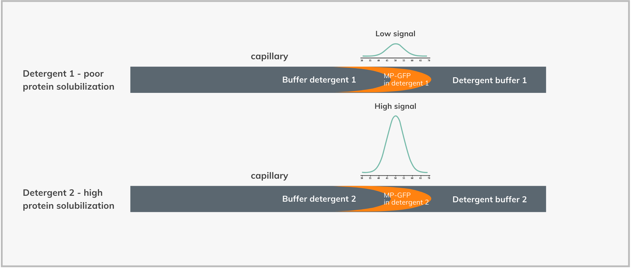
Instrument:
Fida 1 with 480 nm LED fluorescence detection. Fidabio standard capillary (i.d.: 75 μm, LT: 100 cm, Leff: 84 cm). The sample format was Fidabio glass vials for all the tests.
Indicator:
The indicator was the crude lysate containing the solubilized GFP-tagged protein.
Analyte:
The analyte is just the buffer containing the corresponding detergent used to solubilise the protein of interest.
FIDA experiments:
20μL of crude membrane solubilised with a specific detergent was loaded into a Fidabio glass insert. A total of 12 samples, each with a different detergent, were loaded into the Fida 1 system, and the sample tray was kept at 5°C for the duration of the whole measurements. The washing buffers and the capillary chamber were at 25°C. The method shown below was used for the GFP-LacY experiments:
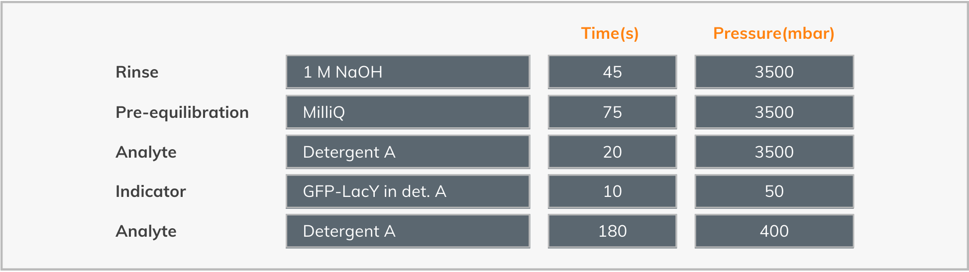
Membrane protein sample preparation:
The cells overexpressing the protein of interest were lysed, centrifuged and the insoluble fraction resuspended in PBS, aliquoted and frozen. Afterwards, a specific detergent buffer was added to a crude membrane aliquot to solubilize the protein of interest. The samples were ultracentrifuged at 100,000 x g for 45 mins at 4 0C. The supernatant containing the solubilized membrane protein was diluted to 0.1 mg/ml of total protein concentration, determined with Bradford assay. The diluted sample was measured with the Fida 1 instrument and FSEC.
Buffer composition: 1X PBS, 150 mM NaCl + detergent. Final detergent concentrations: CHAPS 2%; CHAPSO 2%; CYMAL-6 1%; C12E8 1%; DDM 1%; DM 1%; FC12 1%; LDAO 1%: LMNG 1%; OG 2%; UDM 1%; Control: No Detergent.
RESULTS
Flow-Induced Dispersion Analysis
The GFP-LacY was solubilised in 11 different detergents plus a negative control without any detergent. Figure 3A shows the histograms of the peak area for all detergents, suggesting that DDM and UDM most effectively solubilise GFP- LacY. The raw signals used to calculate the peak areas are overlayed in Figure 3B. Fida 1 also measures the Polydispersity Index (PDI) based on the peak shape. Polydisperse samples have higher PDI values, meaning that the indicator exists in a distribution of different sizes. In contrast, when the PDI is below 0.05, the sample is considered monodisperse.
Among the detergents screened in this assay, CHAPS, CHAPSO and OG have relatively high PDI values, while the rest of the detergents have similar values, close to 0, indicating monodisperse samples (Figure 3C).
These data show that Fida 1 can accurately screen for different detergents in unpurified samples, using only 40 nL per measurement, assessing 12 conditions in triplicates in just 3 hours. Most importantly, the volume used to load the instrument - 20μL - is fully recoverable and can be used for downstream analysis eliminating batch-to batch differences.

A. FIDA detergent screening of 0.1mg/ml of GFP-LacY solubilised in different detergents. GFP-LacY peak area histograms for each detergent,
B. Peak overlay for each of the GFP-LacY signals in different detergents, C. PolyDispersity Index (PDI) calculated for each GFP-LacY signal in different detergents
Fluorescence Size-Exclusion Chromatography
To validate the FIDA data for membrane protein detergent screening, the samples were also analysed with Fluorescence Size-Exclusion Chromatography (FSEC), one of the standard methods for detergent screening. The signal from the FSEC is used to assess the level of solubilised protein in a detergent buffer, as well as the dispersity of the protein of interest. Figure 4A shows the overlapping FSEC chromatograms of all 12 conditions. UDM and DDM were shown to be the best detergents for solubilising GFP-LacY confirming the results obtained with FIDA.
These data demonstrate that the Fida 1 system provides comparable results to the technologies used to date, with the striking advantages of using 10 to 100 times less sample volume, being much faster and offering sample recovery opportunity as summarised in Table 1. Figure 4B compares detergent performance ranking with FSEC and FIDA based on signal intensity, while taking into consideration that the FIDA assay was run in triplicate while FSEC just one replica. The two rankings match very well, given that DDM and UDM have practically the same intensity in the two technologies.

A. Fluorescence Size-Exclusion Chromatography (FSEC) detergent screening of the samples of Figure 3.
B. Screening ranking comparison between the FIDA assay and FSEC, ranking 1 was the best detergent that solubilized the most protein. ** The samples with DDM and UDM have practically the same level of solubilised GFP-LacY.

CONCLUSIONS
The data demonstrate that the Fida 1 system offers a new approach to detergent screening that has significant advantages allowing essential data to be obtained faster using less material. The FIDA assay uses only 120nL of crude protein solution at 0.1 mg/ml for three replicates, and the whole volume leftover (~19.8 μL) is fully recovered, available for further experiments. The experimental time was less than 3 hours for 12 detergents in triplicates compared to ~20h for one replica in FSEC. In addition to FSEC and Fida 1 providing the same answers, Fida 1 gives a quantifiable PDI.
Altogether, the results of this work presents a new approach to characterise membrane proteins that allows users to reducing costs and time as well as to analyse protein expressed at low levels in a short time thus overcoming any stability issues.