
Protein Oligomerization: Easy and Accurate determination in plasma and buffer
INTRODUCTION
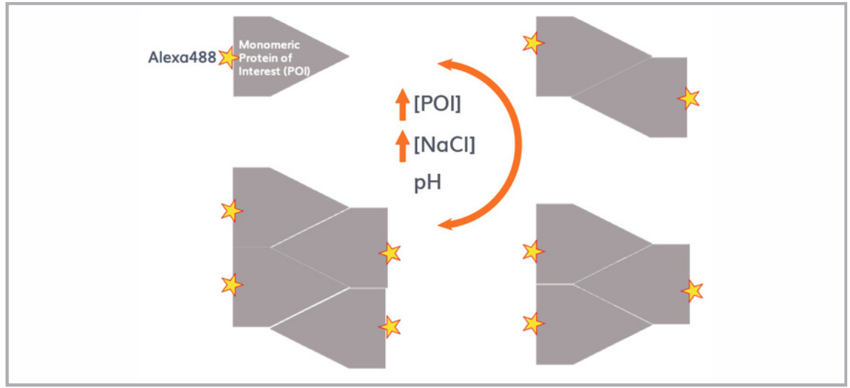
The active state of many proteins, including many drug targets, primarily exists as dimers or oligomers. Understanding the conditions under which oligomerization happens is paramount in drug development, formulation, and bioprocessing. In this work, we have analyzed an injectable therapeutic protein which can appear as monomer, dimer and tetramer; of which only the latter binds to its target receptor. As the protein is active in the bloodstream, we also performed experiments in 90% plasma to simulate physiological conditions. All the experiments were conducted, using a Fida 1 instrument, enabling accurate detection of protein size and size changes under different conditions. A distinctive feature of the Fida 1, is the possibility of analyzing directly in crude matrices such as plasma and serum. In this application note we demonstrate how Fida 1 provides decisive information on protein oligomeric state, not achievable with any other methods. The following assays were realized:
i) Effect of ionic strength
ii) Effect of pH
iii) Sample matrix: Buffer & 90 % plasma
MATERIAL & METHODS
Fida 1 instrument with 480 nm ex LED fluorescence detection for binding experiments. Fidabio standard capillary (i.d.: 75 μm, LT: 100 cm, Leff: 84 cm). Flow-Induced-Dispersion-Analysis was performed by filling the capillary with the analyte (buffer or unlabeled POI), followed by an injection of 40nL of POI-alexa488 or preincubated POI alexa488+POI, which was mobilized towards the detector at 400 mbar.
i) Effect of ionic strength
Acetate phosphate buffer, 0.03% Pluronic Acid F127, pH 5,0. NaCl was titrated from 0,5 to 150 mM. Protein of interest (POI) was used as the indicator at 25 nM, labeled with Alexa Fluor® 488 Protein Labeling Kit from ThermoFisher Scientific.
ii) Effect of pH
Acetate phosphate buffer, 0.03% Pluronic Acid F127, 100 mM NaCl. pH was varied from 3.5 to 7.2 in the pH titrations. Protein of interest (POI) was used as the indicator at 25 nM, labeled with Alexa Fluor® 488 Protein Labeling Kit from ThermoFisher Scientific.
iii) Sample matrix: Buffer & 90 % plasma
Acetate phosphate buffer, 0.03% Pluronic Acid F127, 100 mM NaCl, pH 5,0. POI- Alexa488 was kept constant at 10 nM, and unlabeled-POI was titrated from 10nM to 16μM, using standard buffer or plasma as matrix.
RESULTS
Compared to other fluorescent-based techniques, the Fida 1 provides direct measurements of an absolute value, the diffusion coefficient, which is directly linked to the hydrodynamic radius (Rh).
i) Effect of ionic strength
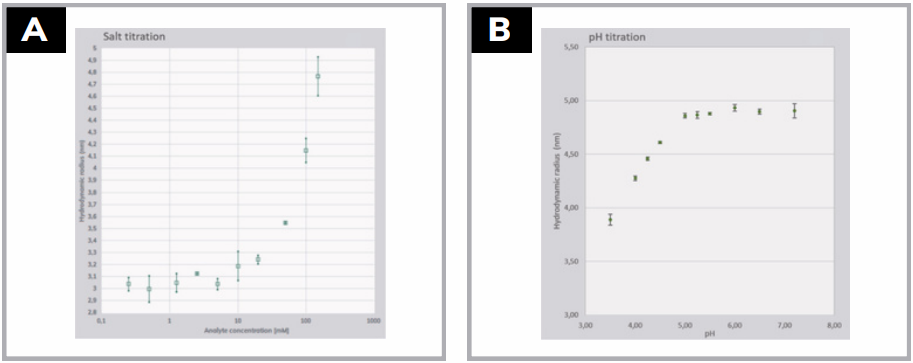
Figure 2A reveals the impact on size with variation in NaCl concentration. Below 10 uM, the size corresponds to that of a monomer. From 10 uM NaCl upwards, the Rh increases from the monomeric size of 3 nm as more and more multimeric forms assemble. The 10 different titration points have an average error of only ±0.077nm, which is between 1-1.5% of the measured Rh.
ii) Effect of pH
Acetate phosphate buffer, 0.03% Pluronic Acid F127, 100 mM NaCl. pH was varied from 3.5 to 7.2 in the pH titrations. Protein of interest (POI) was used as the indicator at 25 nM, labeled with Alexa Fluor® 488 Protein Labeling Kit from ThermoFisher Scientific.
iii) Sample matrix: Buffer & 90 % plasma
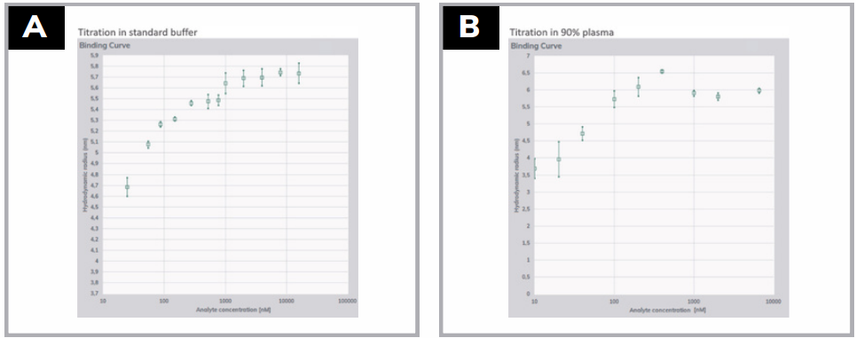
The following sets of experiments were designed to illustrate the use of the Fida 1 for concentration dependence of the oligomeric state both in buffer and in plasma. The optimal pH for observing concentration dependent oligomeric changes was identified to be around pH 5. The concentration dependent formation of the tetramer was detected by titrating the labelled POI with un-labelled POI from 0 to 16 uM. As seen in Figure 3A, the measured size of labelled POI alone was 4,7 nm which increases with increasing amounts of un-labelled POI. At 2 uM unlabeled POI a saturation is reached at 5,7 nm indicating that all labelled POI is on tetrameric form. Error bars in buffer are in the range 1-4%. The data displayed in Figure 3B shows the same assay as 3A, only run in 90% plasma instead of buffer. The Alexa488 labelled POI now displays a Rh of 3,7 nm which is close to monomeric state. The transition towards full oligomerization takes place over a narrower concentration range in plasma vs. buffer, and already at 100 nM is the maximum size reached. Maximum in plasma is around 6,0 nm vs. 5,7 nm in buffer. Error bars in plasma are in the range 1-10%.
CONCLUSIONS
Oligomerization of proteins is known to occur for many systems. However, the impact of environmental parameters such as Ionic strength, pH and sample matrix is notoriously hard to assess using a single methodology. The FIDA technology enables easy and reliable assessment of the oligomeric state of protein drug targets in varying assay conditions. In addition, it can confirm the oligo- meric state of the protein in crude matrices such as plasma thus providing important information on protein’s confirmation in near physiological conditions. Fida 1 therefore can be used to conveniently optimize protein condition for example in formulation studies and bioprocessing routines.