Reversible oligomerization and FIDA: From label free detection to oligomerization Kd determination.
The active state of many proteins are homo-oligomers, this includes several drug targets such as the HIV-1 protease, EGFR, HER2 and TNFα among many others.1–3 The oligomeric state of a given drug targets has massive implications on designing binders against it as it can impact the stoichiometry, avidity effects and stability in solution.3
In this app note we present how to detect and characterize homo-oligomerization using Flow Induced Dispersion Analysis (FIDA) in a label free manner. FIDA allows sub angstrom scale precision in determination of the hydrodynamic radius (Rh),4 this combined with a measurement range from ~500 nM to mM protein concentration allows the method to be an extremely versatile tool for both detection and characterization of homo oligomers.
Workflow and example results
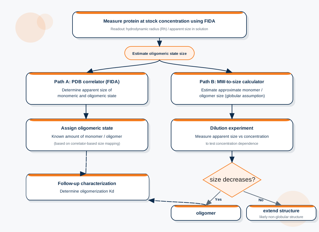
As homo oligomerization is a protein-protein interaction it is concentration dependent. Hence, it is recommended to measure the Rh at the stock concentration of the protein (Figure 1). The Fida instrument only consumes 40 nL of the loaded amount into the autosampler. The remaining sample can either be recovered for parallel experimentation or used for subsequent characterization.
To demonstrate the workflow here we characterize the homo oligomerization of bovine β-lactoglobulin (BLG) a well known homo-dimer which forms pH and salt dependent higher order oligomers.5
Step 1: Measure the protein at stock concentration
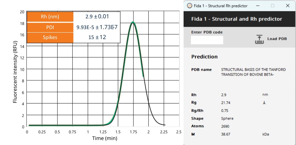
1096 µM of BLG in PBS pH 7.4 + 150 mM was measured at 25°C. The protein is highly monodispersed (PDI < 0.05) and contains a low number of spikes considering the mM concentration. Symmetrical Taylorgram indicated no stickiness. Overall, the sample QC suggest a high-quality sample.
Step 2a: PDB correlator-based determination of oligomeric state
If PDB models from either ALFA-fold or the protein data bank are available, the PBD correlator in the FIDA software can be used to predict the Rh of the protein. Using a dimeric crystal structure of BLG at neutral pH (1BSY) as well as the monomer structure the Rh was estimated (Figure 2).
Step 2b: molecular weight-based approach
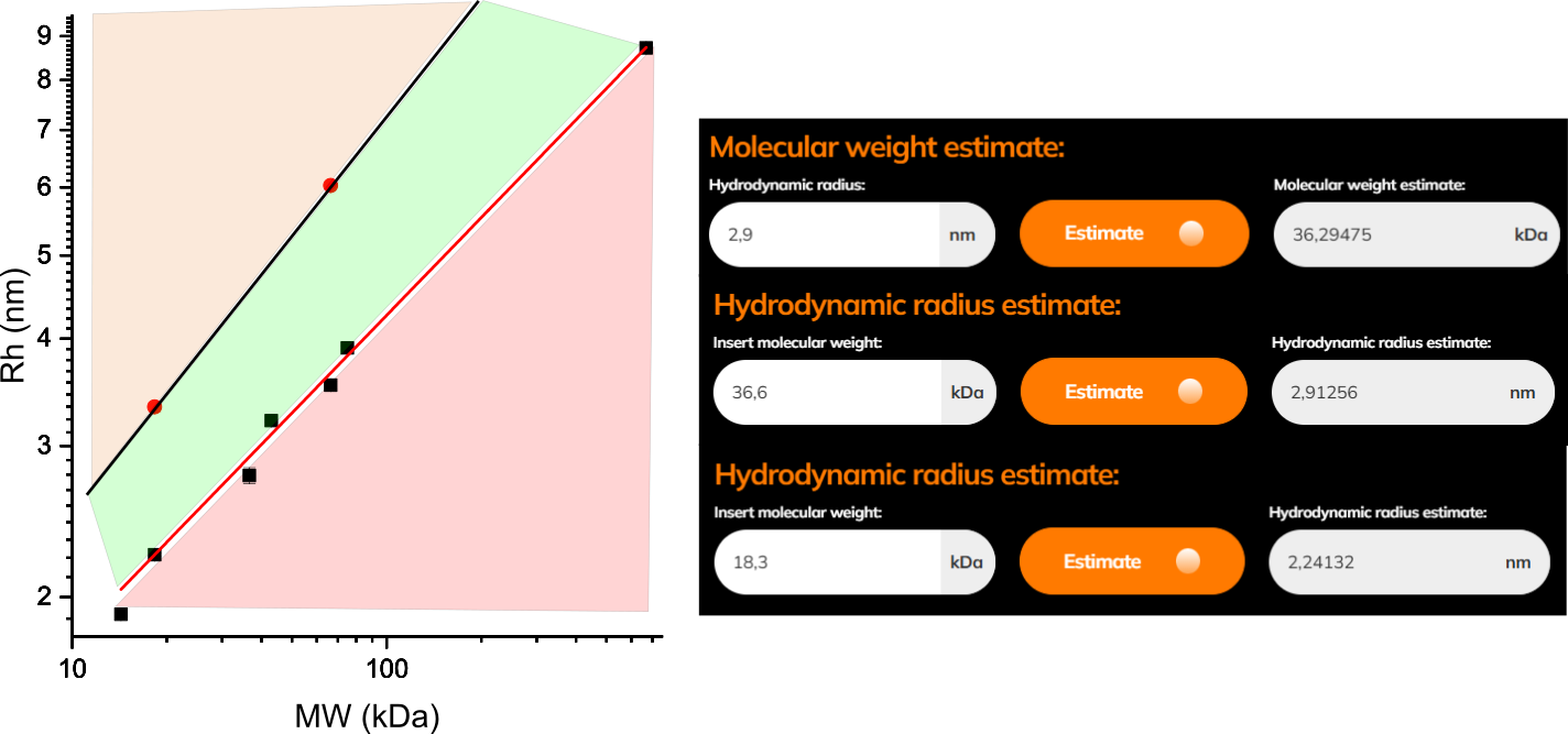
Assuming globularity the Rh can be predicted based on the molecular weight (MW) of a given protein and vice versa (Figure 3). Using the Rh-to-molecular weight converter (molecular-weight-to-size-calculator), the measured size of BLG i.e., 2.9 ± 0.01 nm corresponds to 36.3 kDa. The reported molecular weight of BLG is 36.6 kDa, which confirms dimeric state of BLG at measured concentration.
NOTE: proteins that are not globular may have an apparent Rh larger than that expected for a globular protein and thus will lie in the green zone of the illustrated Rh-to-molecular weight correlation curve (Figure 3).
To alleviate this challenge an experimental proof of oligomerization can be performed.
Step 3: dilution experiment
Table 1 – Measured Rh values for BLG at stock and diluted concentrations. The reduction in Rh upon dilution confirms concentration-dependent dissociation consistent with reversible oligomerization.
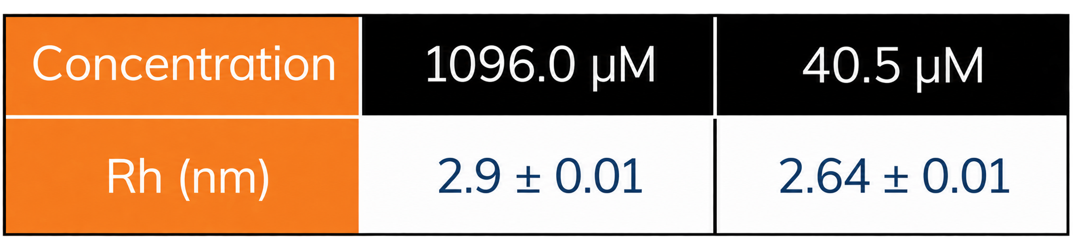
Using the same sample from above measurements to perform a dilution point and measuring the apparent Rh of the new sample yielded a smaller Rh (table 1), as all non-covalent oligomerization is protein concentration dependent, this indicates oligomer dissociation. This approach enabled confirmation of protein oligomerization with a total instrument time of 10 min.
Detailed characterization option 1: The influence of sample matrix
FIDA is largely independent of sample matrix composition, enabling measurements to be performed under a wide range of physiologically relevant conditions. Using UV detection, the effects of factors such as pH, ionic strength, detergents, and other buffer components on protein size and conformational state can be evaluated directly. Furthermore, fluorescent labelling of the protein of interest enables the investigation of protein oligomerization and intermolecular interactions in complex biological matrices, including cell lysates and serum.6
To demonstrate this, the oligomeric state of BLG at several different pH was assessed labelfree (Figure 4).
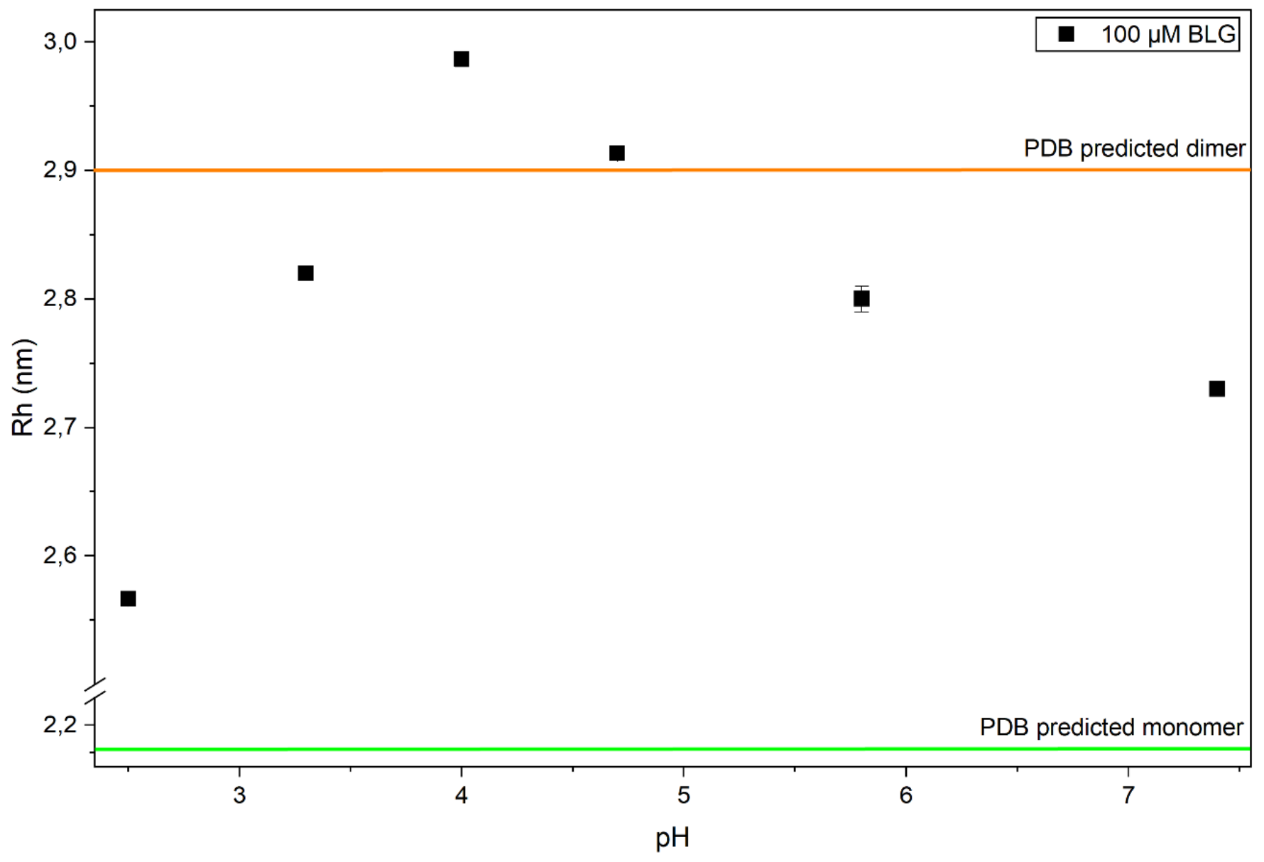
At neutral pH, the protein is not fully dimeric even at a concentration of 100 µM. The propensity for dimerization increases at pH 4.8, whereas at pH values below 3 the monomeric form becomes predominant. Around pH 4, the formation of higher-order oligomeric species is observed. These findings raise important questions regarding the stability of the oligomeric species and the equilibrium constants governing the oligomerization process.
Detailed characterization option 2: Determination of oligomerization Kd
Determination of the equilibrium dissociation constant (Kd) for protein homo-oligomerization is straightforward using FIDA. A concentration series of the protein of interest is prepared and analyzed using the complex dissociation (CapDis) workflow, allowing the oligomerization equilibrium to be monitored directly and the corresponding Kd to be determined (Figure 5).
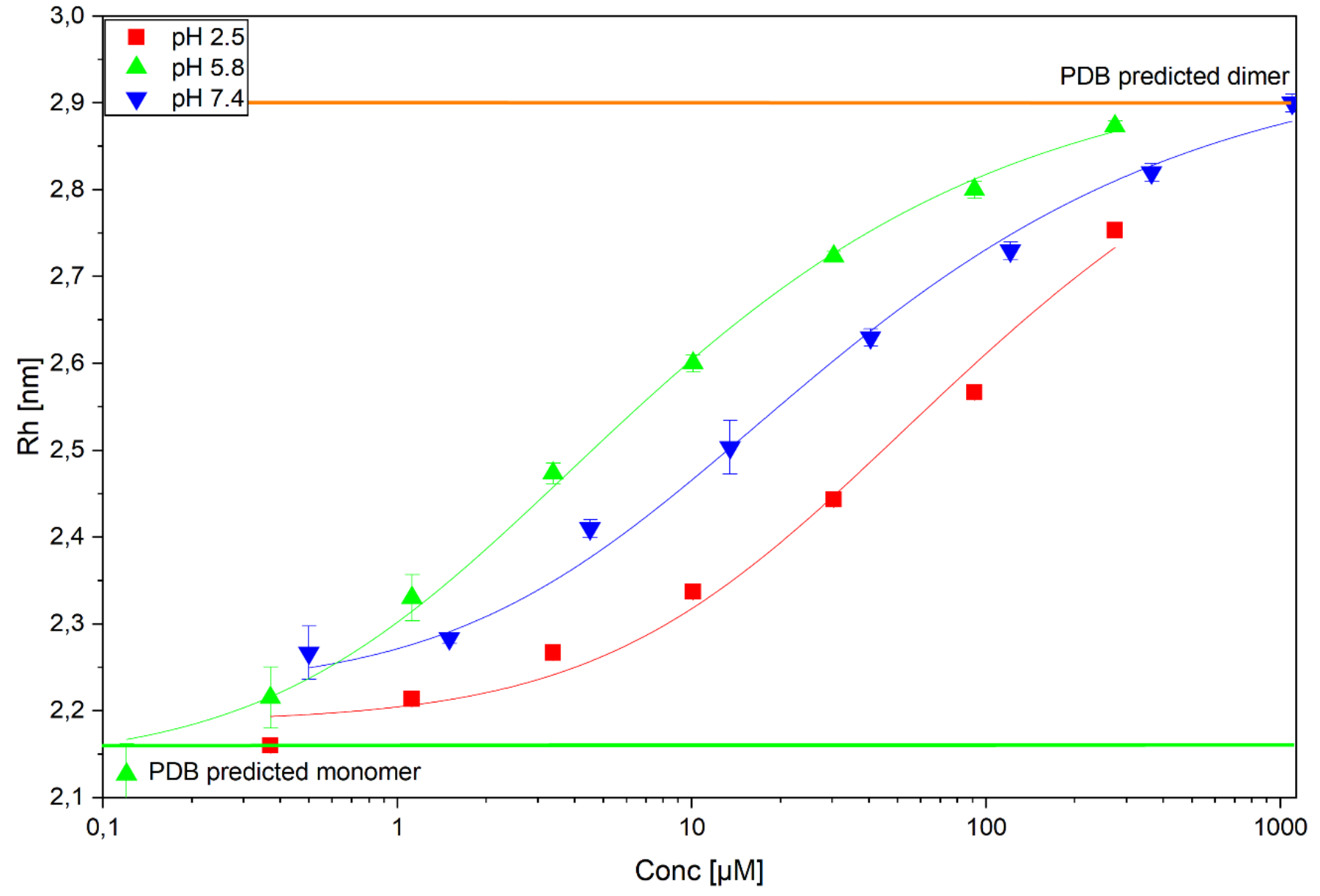
The oligomerization curves obtained may not fall into the standard protein-protein binding curve analysis assumptions but can be modelled using a specific modelling approach. A description of model used in the current study is provided under the appendix section of this application note.
Table 2 – Oligomerization parameters across pH conditions. Fitted dissociation constants (Kd) and extracted species radii obtained from concentration-dependent FIDA measurements at different pH values.
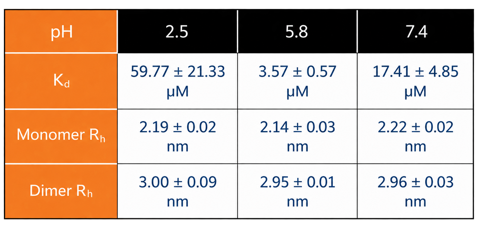
As we lower the pH from neutral pH the dimer becomes more favoured. At low pH the monomer is favoured but the dimer is still present.
Conclusion
Flow Induced Dispersion Analysis (FIDA) provides a robust approach for studying protein homo-oligomerization by directly measuring hydrodynamic radius in solution with high precision. Because the measurement is performed under native conditions without immobilization or surface attachment, the method enables characterization of self-association equilibria with minimal perturbation to the system. Across a broad concentration range, from sub-micromolar to millimolar protein concentrations, subtle concentration-dependent changes in hydrodynamic radius can be resolved, allowing oligomer formation to be monitored directly.
Unlike surface-based or intensity-dependent techniques, FIDA provides population-averaged hydrodynamic size measurements that can be readily interpreted using thermodynamic models. As demonstrated here, concentration-dependent self-association can be identified through simple dilution experiments, while quantitative analysis of complete concentration series enables determination of oligomerization equilibrium constants.
In addition to its analytical capabilities, FIDA offers several practical advantages, including low sample consumption, short analysis times, and compatibility with a wide range of buffer compositions. When combined with fluorescent labeling, the technique can also be applied to complex biological matrices such as serum and cell lysates. Together, these features make FIDA a versatile platform for investigating protein homo-oligomerization under physiologically relevant conditions.
References
- Todd, M. J., Semo, N. & Freire, E. The Structural Stability of the HIV-1 Protease. J. Mol. Biol. 475–488 (1998) doi:10.1006/jmbi.1998.2090.
- Cheng, X. A Comprehensive Review of HER2 in Cancer Biology and Therapeutics. Genes vol. 15 Preprint at https://doi.org/10.3390/genes15070903 (2024).
- Pedersen, M. E., Haegebaert, R. M. S., Østergaard, J. & Jensen, H. Size-based characterization of adalimumab and TNF-α interactions using flow induced dispersion analysis: assessment of avidity-stabilized multiple bound species. Sci. Rep. 11, (2021).
- O’Dea, F. et al. Improvement in protein HSQC spectra from addition of betaine. J. Biomol. NMR 79, 155–162 (2025).
- Gottschalk, M., Nilsson, H., Roos, H. & Halle, B. Protein self‐association in solution: The bovine β ‐lactoglobulin dimer and octamer . Protein Science 12, 2404–2411 (2003).
- Willmer, P. et al. In-Solution Characterization of Biomolecular Interaction Kinetics under Native Conditions. Anal. Chem. 97, 19498–19504 (2025).