
Selective quantification of IgG Directly in F12 Fermentation Media
Introduction
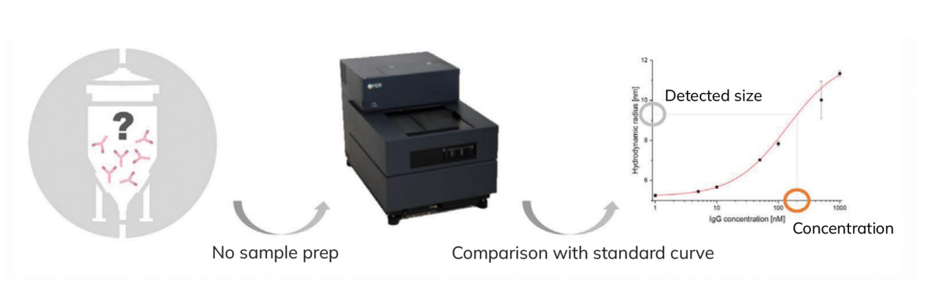
A complex sample matrix still presents a significant challenge in most biomolecular binding assays, thus requiring several purification steps for obtaining reliable assay results. This leads to a long assay development time, a need for time-consuming purification processes, and ultimately an increase in operational costs. Here we present a Flow-Induced Dispersion Analysis (FIDA)-based procedure for quantifying IgG directly in F12 fermentation media, without any need for purification.
FIDA is a new capillary-based technology for quantifying biomolecular concentration directly in complex solutions (e.g. plasma) and for measuring in-solution binding under native conditions. FIDA is based on Taylor dispersion in a pressure-driven flow of a ligand (termed indicator, e.g. Protein-A) interacting with the analyte of interest (e.g. an IgG antibody). The indicator alone appears small (i.e. it has a small hydrodynamic radius) when it is not bound to the antibody, but upon binding it will appear larger (i.e. the complex has a larger hydrodynamic radius). The change in apparent size forms the basis for an accurate measure of analyte concentration and interaction.
Material & Methods
Fida 1 instrument with 480 nm LED fluorescence detection (Fida Technologies ApS). Fida coated capillary (i.d.: 75 µm, LT: 100 cm, Leff: 88 cm). 67 mM phosphate buffer pH 7.4 was used as working buffer, Protein-A-Alexa488 as an indicator (100 nM), IgG from human serum was spiked (0-1000 nM) into 80 % F12/DMEM fermentation media and used as analyte. Sample analysis was performed by filling the capillary with the analyte, followed by an injection of 39 nL indicator, which was mobilized towards the detector with analyte at 400 mbar.
Results
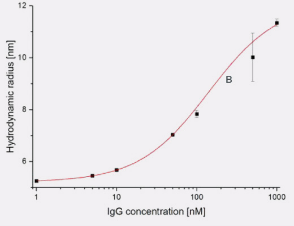
The apparent size (hydrodynamic radius) of Protein-A was plotted as a function of IgG concentration (see figure 1). It was observed that an increase in IgG concentration led to an increase in the apparent size of Protein-A. The unbound Protein-A had a size of 5.5 nm, but upon binding to one or two IgG molecules it increased gradually to approx. 11 nm.
A FIDA binding isotherm was fitted to the obtained data (red line in Figure 1), and the dissociation constant (Kd) for the protein-A-IgG complex in 80 % F12 fermentation media was estimated to be 60 nM. Furthermore, the binding curve can be used to quantify IgG in F12 fermentation media.
Conclusion
The FIDA methodology is applicable for biomolecular quantification and for measuring binding affinity directly in complex samples, thereby reducing sample purification requirements and assay development time.