
Targeted Protein Degradation - Full Characterization of Ternary complexes
VERSION 1.3
Key Benefits of Fidabio in BiDACTM Development:
- Size and Integrity of the Protein of Interest and E3 ligase
- Ternary Complex Binding Affinities
- Fraction of POI bound in Ternary Complex
This application note is developed in close collaboration with C4 Therapeutics, Inc., who is pioneering a new class of small molecule drugs that selectively destroy disease-causing proteins via degradation using the innate machinery of the cell.

INTRODUCTION
When the concept of targeted protein degradation (TPD) was first introduced 20 years ago, it was considered a curious academic exercise (1). Since then, TPD has evolved into one of the most promising therapeutic modalities gathering broad scientific interest and significant investments.
Contrary to standard inhibiting drugs, “degraders” trigger an endogenous cell mechanism, targeted protein degradation, which captures and “slays” unwanted or damaged proteins via the ubiquitin- proteasome system.
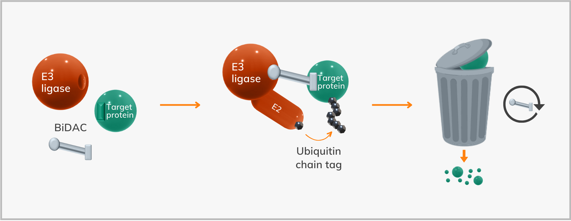
Degraders or BiDACTM (Bifunctional Degradation Activating Compounds) molecules, are bifunctional compounds composed of three parts: One that binds to the protein of interest, a linker, and a moiety that binds to the E3 ligase; together forming a ternary complex bringing the E3 ligase and the Protein of Interest (POI) in proximity, whereby ubiquitin may bind to the latter and subsequently the POI is degraded by the proteasome.
Characterization, stability, and formation of the ternary complex are critical components in designing the best drug candidates (2).
In this application note, in collaboration with C4 Therapeutics, Inc., we show how the Fida 1 can provide a full characterization of the ternary complex and its subunits, including Kds of all binding partners, sizes of subunits and the ternary complex, as well as simple integrity control.
MATERIAL AND METHODS
Fida 1 instrument with 480 nm LED fluorescence detection for binding experiments (Fida Biosystems ApS). Fida standard capillary (i.d.: 75 μm, LT: 100 cm, Leff: 84 cm). HEPES buffer 10mM pH 7.4, 200mM NaCl, 0.05% Pluronic acid F127, 1mM TCEP was used as the working buffer.
The POIs were used as the indicator, labeled with Alexa Fluor® 488 Protein Labeling Kit from ThermoFisher Scientific. The POIs in this system will be called POI1 and POI2. A titration of the BiDAC molecule was performed where the POI and E3 ligase were kept constant at 10nM and 40nM respectively and four different BiDAC molecules have been titrated as analytes from 0-10μM. Flow-Induced-Dispersion-Analysis was performed by injecting the capillary with the analyte + E3 (4μL), followed by an injection of 39 nL of preincubated POI+analyte+E3, which was mobilized towards the detector at 400 mbar.
RESULTS
Quantitative determination of ternary complex and its subunits
Compared to TR-FRET, AlphaLISA, and other proximity ligand-binding assays, FIDA provides direct measurements of an absolute value, the hydrodynamic radius (Rh). I.e., it is calibration-free, and risks of false positive/negative are eliminated.
Before the BiDAC titration, the size of the individual POI, the POI in the presence of E3, and the unlabeled E3 were measured. The Rhs of labeled POI1 and POI2 alone were 2.85 and 2.51 nm respectively. As a control, and to test potential positive cooperativity, the POI in the presence of E3 was analyzed showing a size identical to the POI alone, indicating that the POI and the E3 would not interact in the absence of the BiDAC.
To verify the size of the unlabeled E3, intrinsic fluorescence detection was performed on the Fida 1 showing a Rh of the E3 of 5.8nm. Subsequently, four different BiDACs, P1, P2, P3, and P4 were titrated against POI1 and four BiDACs P5, P6, P7, and P8 against POI2. When three components are involved in a binding, as for the ternary coplex, a bell-shaped binding curve is theoretically predicted as a function of the BiDAC concentration (Figure 2).
With increased concentration of the BiDAC, it is observed that ternary complexes shift to binary complexes (2,3). Here Fida 1 monitors the size of the labeled POI, and the titrations of BiDACs display the predicted size increase at lower concentrations followed by a size decrease at higher concentrations (Figure 3), as predicted by the models in the literature (2,3).
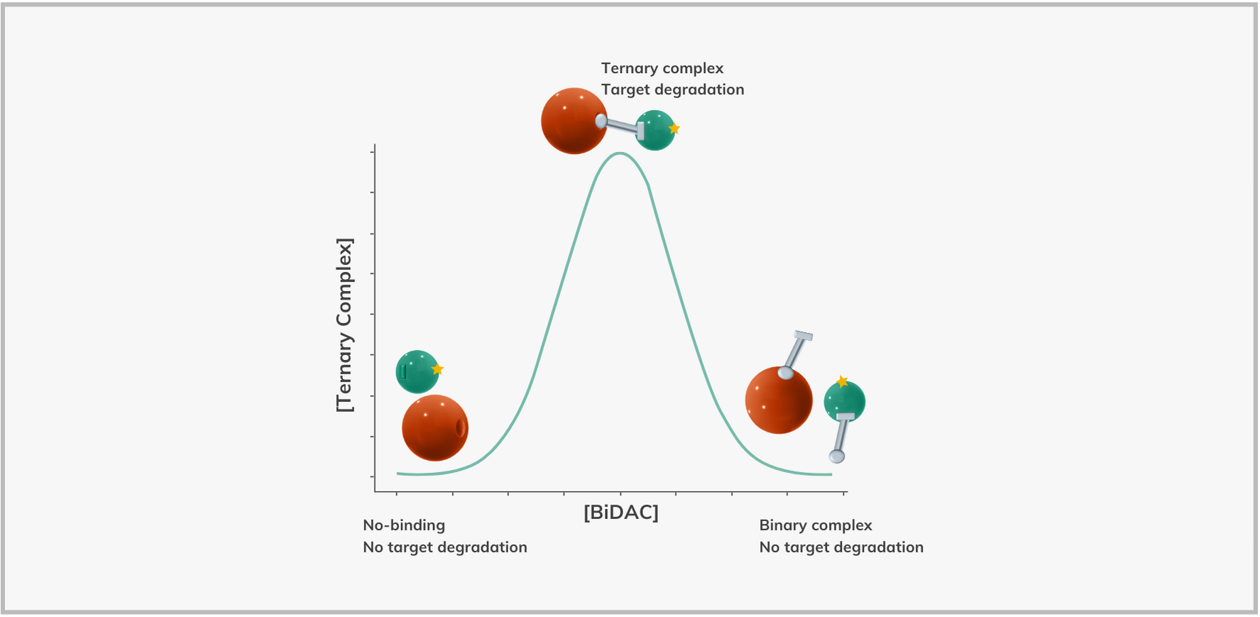
E3 ligase (orange), POI (mint green), fluorescent label (star), BiDAC (silver-bar).
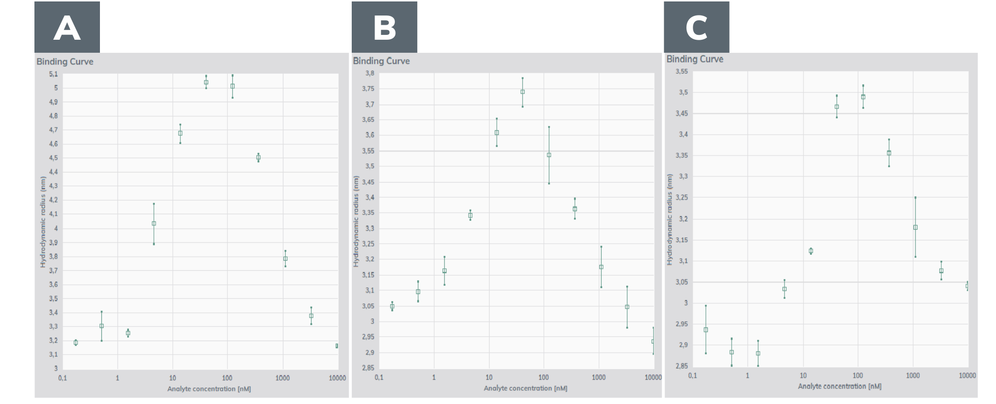
POI 10nM, E3 40nM, BiDAC titrated from 0-10μM. (A) POI1 + E3+ P3; (B) POI1 + E3 +P4; (C) POI2 + E3 +P6.
Figure 3 shows the data of the titrations, where the concentration of the POI and the E3 were kept constant, and the different BiDACs were titrated. The binding curves have the bell-shape as predicted by the models. Compared to relative technologies, Fida 1 measures an absolute parameter (size), and the size increase is directly proportional to the amount of ternary complex. Thereby, Fida 1 allows you also to assess the fraction of the POI bound in the ternary complex.
FIDABIO SIMULATION MODEL
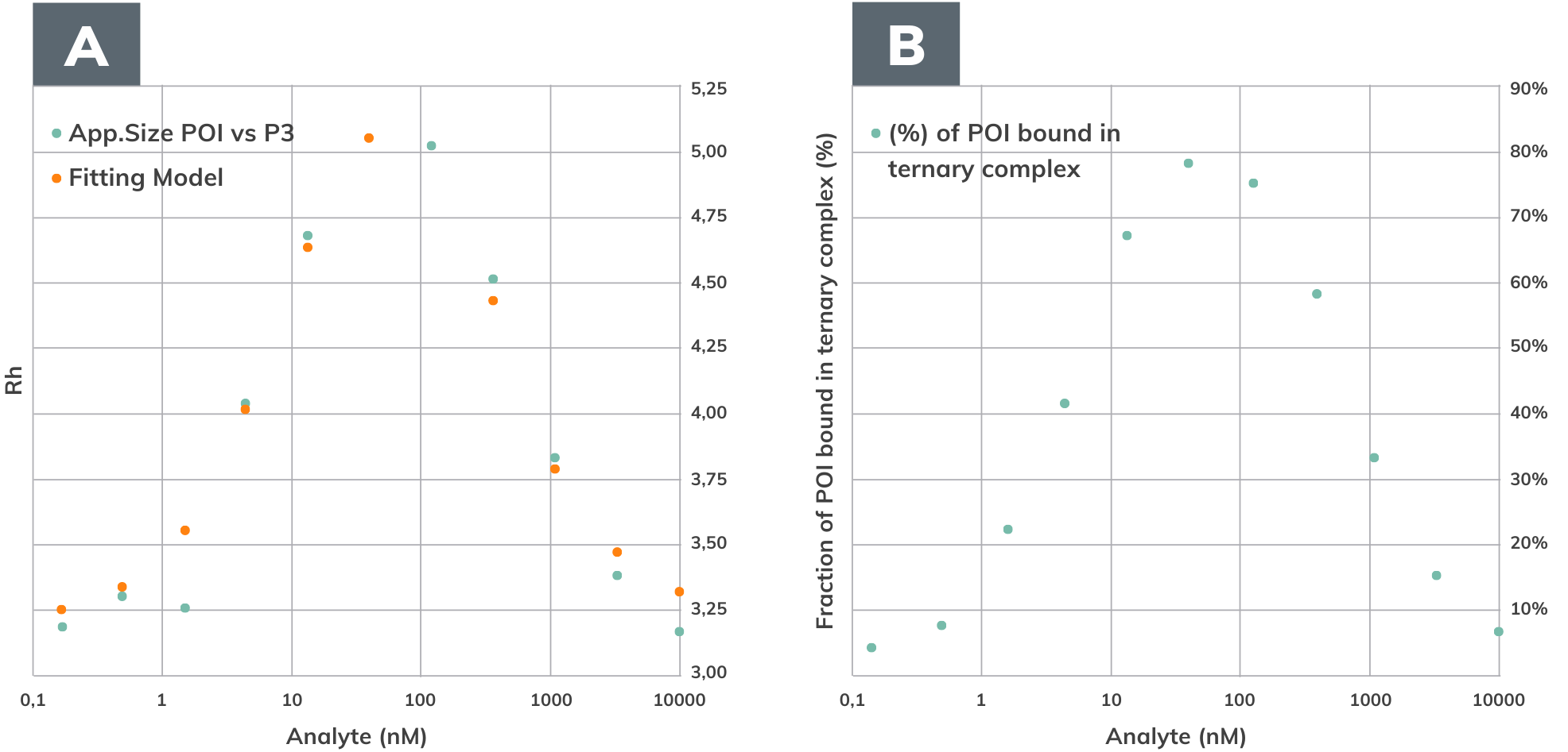
As the ternary complex involves multiple binding affinities, standard binary fitting equations do not apply. Therefore, Fida Biosystems has developed a model to simulate a specific ternary complex binding based on the size of the POI, the POI and the BiDAC and the E3. By knowing the diffusion coefficient and the Rh of these components, we can calculate the fraction of POI bound in the ternary complex.
In this case the apparent diffusion constant is described as follows:

where x is the fraction of PDE, (the ternary complex), y is the fraction of PD (the POI and the BIDAC), z is the fraction of P (the POI); DifPDE, DifPD, and DifP are the diffusion coefficients of PDE, PD and P respectively. The cooperativity alpha is also taken into account in the model, with negative cooperativity having alpha <1 and positive cooperativity alpha >1 (2).
The apparent hydrodynamic radius (Rapp) is determined in the Fida 1, and under these conditions it is expressed as follows:

I.e., with the fitting model shown in Figure 4, we can calculate: Cooperativity, dissociation constant between the POI and the BIDAC (Kd[PD]), dissociation constant between the BIDAC and the E3 (Kd[DE]), the ternary complex size, and the fraction of POI bound in the ternary complex. The specific values are reported in the Figure 4 legend.
Fida 1 sets a new standard for characterization of BIDACs that will allow users to get a full picture of the drug candidate. Please contact us for further mathematical details on the model.
CONCLUSIONS
The Fida 1 provides an opportunity for fast and quantitative determination of ternary complex conformation and accurate determination of the binding interactions between the various subunits under different conditions. Combined with low sample consumption, easy sample preparation and a comprehensive, user-friendly software suite, Fidabio offers a new technology for characterizing complex modalities, such as BiDACs, allowing the users to get full insight of ternary complex formation as well as other essential parameters in BiDAC development. Additionally, the size of individual proteins gives precise information about the integrity and stability of the different partners.
As an additional step in the BiDAC development, Fida 1 is also able to detect in-vitro ubiquitination without the tedious and time-demanding work of SDS-page and western-blot. Please visit our website for the application note on ubiquitination.
REFERENCES
1. Sakamoto KM, Kim KB, Kumagai A, Mercurio F, Crews CM, Deshaies RJ. BiDACs: Chimeric molecules that target proteins to the Skp1-Cullin-F box complex for ubiquitination and degradation. Proc Natl Acad Sci U S A. 2001;98(15):8554–9
2. S. L. Fisher, A. J. Phillips, Targeted protein degradation and the enzymology of degraders. Curr Opin Chem Biol 2018, 44, 47–55.
3. Douglass EF, Miller CJ, Sparer G, Shapiro H, Spiegel DA. A comprehensive mathematical model for three-body binding equilibria. J Am Chem Soc. 2013;135(16):6092–9.