
How can you measure amyloid fibril length directly in cerebrospinal fluid or blood?
How do you recognise a neurodegenerative disease years before it is diagnosed, and then track how fast it is progressing? One academic spin-out’s answer is to stop counting how much of a disease protein is present and instead measure how long its amyloid fibrils have grown, directly in a small volume of cerebrospinal fluid or blood.
That approach is the work of NeurotidalDx (Neurotidal Diagnostics), a spin-out co-founded by Francoise Dekker, Ph.D., in which a fundamental question from her PhD grew into a start-up building an early-detection biomarker for Parkinson's disease. The measurement itself rests on Flow Induced Dispersion Analysis (FIDA), the in-solution technique invented by Fidabio’s founder Henrik Jensen.
You can also watch the full webinar recording.
Why is Parkinson's usually caught so late?
By the time a movement disorder like Parkinson's or a memory disorder like Alzheimer's is diagnosed, the disease is already well advanced. The timeline begins roughly 20 years before diagnosis, when the underlying biology has quietly started to go wrong, and only surfaces as symptoms much later.
Two problems follow. First, at the moment of diagnosis, the zero point on that timeline, an estimated 50 to 60% of the brain damage has already been done, so an intervention at that stage arrives late. Second, symptoms are a noisy ruler: they fluctuate day to day, so a patient can have a good day at one clinic visit and a bad day at the next, which says little about the true stage of the disease.
“And therefore we need new, objective biomarkers,” Francoise Dekker said.
What are amyloid fibrils, and why measure their length?
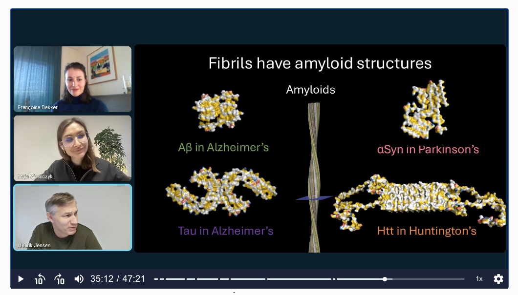
Neurodegenerative diseases share a common feature: amyloid fibrils, ordered clumps of protein that accumulate in the brain before cells die. A healthy, functional monomer unfolds, exposes its vulnerable regions, and other copies stack on top over and over, producing a long, highly repetitive fibril. Different diseases feature different proteins, tau and beta-amyloid in Alzheimer's, alpha-synuclein in Parkinson's, and huntingtin in Huntington's, but all of them stack into the same kind of elongated structure. The ordered fibril cores come from high-resolution cryo-electron microscopy and recur across patients with the same disease.
Dekker's academic group studied a very fundamental question: how these fibrils grow, and how the cell's own machinery, a family of proteins called chaperones, tries to slow them down or break them apart. A concern sits underneath that question: if disaggregation is incomplete, you can end up with many more fibrils, each with new growing ends, potentially accelerating the disease. To study any of this properly, the group needed a tool that could measure fibril length.
The discovery: a label that lights up only fibrils
The team first invested in a FIDA instrument because they saw its potential to measure fibril length in solution. FIDA is an in-solution technique that reports the hydrodynamic radius, the effective molecular size, of whatever a fluorescent probe is attached to. The missing piece was a way to put a fluorescent tag specifically on amyloid fibrils.
After a few years of work, that piece became a compound the team calls FibrilPaint. On its own, the label is small and disperses narrowly, giving a small measured size. When it binds a fibril, the peak broadens and the measured size jumps, and crucially, the longer the fibril, the broader the peak. In effect, FIDA plus FibrilPaint turns fibril length into a readout.
An early tau dataset showed the selectivity. FibrilPaint alone read small; adding healthy monomer produced no change; adding fibrils produced a large increase in size.
“So we only measure the disease-relevant particle,” Francoise Dekker said.
Repeated across time points, the same measurement traces a growth curve, fibrils getting longer and longer, detectable from the earliest aggregates just a few nanometres in size. The information is both qualitative and quantitative: FIDA detects that fibrils are present and measures how long they are, and the balance between free and bound label even gives a sense of how much is there. In her terms, detection is seeing that something is binding, while monitoring is measuring the length and how it changes over time.
From a lab tool to a biomarker in blood and spinal fluid
While developing the method, the team noticed a broader opportunity. A wave of publications pointed to three things: amyloid fibrils leak out of the brain into biofluids such as cerebrospinal fluid and blood; they appear early, before symptoms; and their length tracks how far the disease has progressed.
That reframed the disease timeline. If fibrils show up in blood and spinal fluid roughly 15 to 20 years before diagnosis and then lengthen as the disease advances, measuring their length could both flag the disease early and indicate its stage, the objective ruler that symptom scoring cannot provide. Dekker was careful about the mechanism: the fibrils are not extracted from the brain but leak out on their own, and how exactly that happens is still debated.
Why FIDA works directly on patient samples
A defining strength of the approach is how little sample preparation it needs. Because FibrilPaint is selective and FIDA reads out in solution, the team can measure a patient sample, spinal fluid or blood, essentially as-is, with very little background to subtract. That is part of why others had struggled to turn fibril length into a biomarker: isolating single fibrils by microscopy simply does not scale.
“We always request 100 microlitres, but we only use 10,” Francoise Dekker said.
Volume is modest, a small subset of what a clinical study already collects, so no extra puncture is needed. A subtle but important design choice matters for early detection, where disease proteins are scarce and dilute in blood: NeurotidalDx bases its readout on fibril length rather than fibril concentration.
“We do not use the concentration as a readout, but the fibril length,” Francoise Dekker said.
Concentration-based assays such as ELISAs often show large patient-to-patient and method-related variation; anchoring the measurement to a single, direct size interaction sidesteps much of that. The team is still defining the method's detection limits, and notes it can already bind very early species, fibrils only a few protein layers long.
From PhD project to start-up: building NeurotidalDx
Turning a measurement into impact for patients was a separate journey. The idea began during Dekker's PhD, and few researchers spin a company out of their doctoral work. From there the team brainstormed applications, validated them with doctors, investors and other stakeholders, won early feasibility funding to test proofs of concept, and then raised a first round to work toward an implemented biomarker. In healthcare, the road from proof of concept to market is long, largely because of regulation.
The commercial focus is deliberately narrow at first: biomarkers for Parkinson's disease, a field with fewer tools than Alzheimer's, entering through clinical trials as an exploratory (research-use-only) biomarker where no clinical decision for an individual patient depends on the readout yet. In that setting NeurotidalDx aims to offer three readouts, measuring disease progression more objectively, stratifying patients into groups, and identifying patients earlier. Partners include Pro-Park, the Dutch Parkinson consortium supplying samples for a retrospective study, alongside the Utrecht Incubator community and IP and regulatory advisors.
What's next for early detection of neurodegeneration?
The stakes are large. One widely cited projection holds that by 2050, one in four people will develop a neurodegenerative disease, and there is still no cure. Her framing is that better, earlier, objective measurement is a prerequisite for developing treatments at all.
“We want to be a new standard in neurodegeneration, to replace symptomatic evaluation, enable earlier detection and eventually help therapeutic development,” Francoise Dekker said.
Henrik Jensen sees it as an innovative use of FIDA, with real potential to support the development of new cures for neurodegenerative diseases. It is early, regulation means years of work ahead, but the direction is set: start with Parkinson's, expand to other diseases, and aim for accessible, objective diagnostics of neurodegeneration.
This is the company side of the story. The same FibrilPaint-plus-FIDA approach, used in the laboratory as a molecular ruler for amyloid fibril size and length, is described from the academic side in our conversation with Prof. Stefan Rüdiger.
Curious how FIDA measures size, aggregation and binding in solution? Explore the FIDA technology, see the Fida instrument, learn how FIDA characterises protein aggregation, or browse the peer-reviewed literature. You can also watch the full webinar with Francoise Dekker.
.png)

.png)