
Rapid Sizing And Characterization of Bispecific Antibodies using nL of Sample
INTRODUCTION
Bispecific antibodies are a promising new avenue for immunotherapy treatment of diseases like Multiple Myeloma. They are typically based on monoclonal antibodies with arms that target different antigens on the surface of different cellular targets
Producing the target antigens of bispecific antibodies in-vitro is often costly, time consuming and with low yields. The scarcity of these antigens makes in-vitro optimization of bispecific antibodies a difficult process as scientists are called to extract information from very small volumes.
Here we present how Fida 1 characterizes the formation of ternary complex of bispecific antibodies towards their target antigens using only nL of sample (Figure 1A). Furthermore, using only μL volumes the technology does full titrations yielding the affinity of both antigens, the binary complex size, the ternary complex size as well as the cooperativity (Figure 1B).
The cooperativity is a parameter of utmost importance as strong negative values would disqualify any bispecific antibody for use as a therapeutic
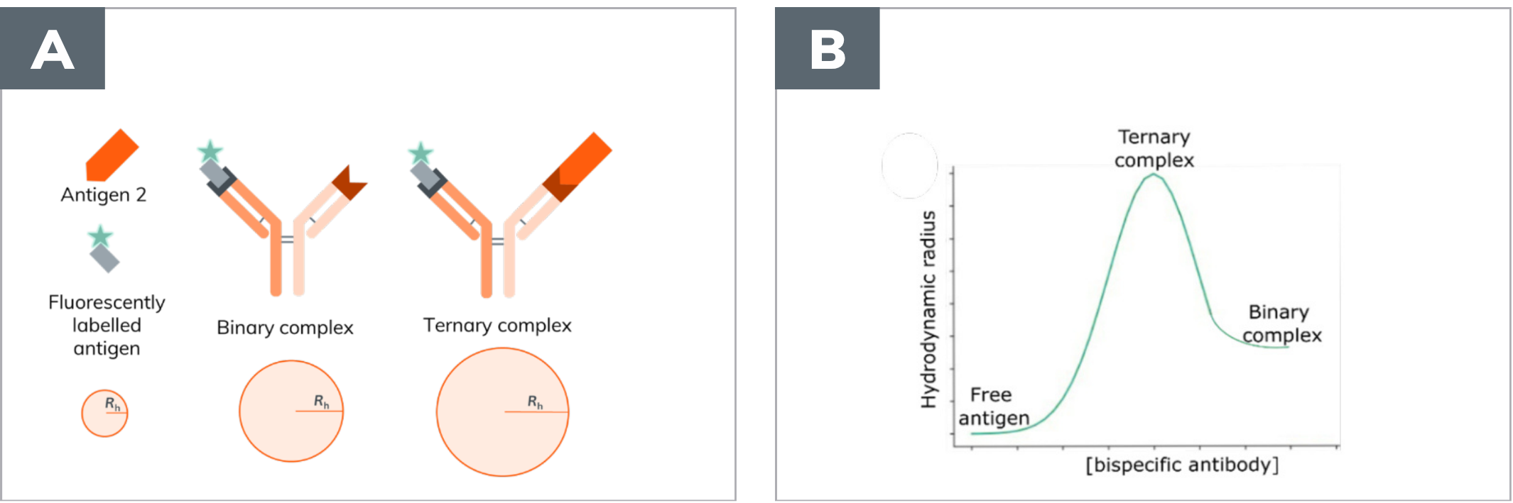
A. Rapid characterization of the size of free antigen, binary and ternary complex using only 120 nL for atriplicate measurement.
B. Full characterization of all binding parameters in the ternary complex formation, including size of all complexes, both K s, as well as the cooperativity, using μL of sample.
MATERIAL & METHODS
Fida 1 equipped with a 480 nm LED fluorescence detector. A Fida coated capillary (L: 1 m, ID: 75μm, L: 84 cm). The buffer used was PBS + 0.1 % BSA for all experiments. Indicator concentration was 37 nM. Unlabelled antigen present in excess.
For ternary complex confirmation, the experiment was run in a capillary dissociation setup: capillary filled with buffer, followed by a 40 nL injection of sample, which was mobilized with buffer.
The full titration was done as a capillary mixing experiment with the capillary filled with antibody (0-2000 nM) and Ag1 (80 nM). A plug injection of labelled Ag2 (37 nM) followed and, finally, the mobilization was carried out with the mixture of antibody and Ag1.
RESULTS
Sixty individual bispecific antibody samples were analysed within 12 hours, using 40 nL of sample per sizing, with no need for user input.
To confirm that the overall architecture of the bispecific antibody has not inhibited the ability to form the ternary complex, all constructs were screened with either labelled antigen.
The positive IgG controls cause a change in hydrodynamic radius upon binding to the labelled target antigen. The same is observed for the fragments specific for Ag1 and Ag2 used to build the bispecific antibodies (Figure 2).
Overall, none of the architectures was found to inhibit ternary complex formation, as a size increase is observed for all cases of binary and ternary complex formation.
Notably, the bispecific antibodies that has multiple fragments specific for the same antigen doesn't cause a further size increase, indicating that extra binding site for each antigen is not beneficial (Figure 2).
.png)
A. Hydrodynamic radius of Ag1 free, in binary (grey) or ternary complex (orange).
B. Hydrodynamic radius of Ag2 free, in binary (grey) or ternary complex (orange).
Total material used per data point is 120 nL for a triplicate.
60 different samples were screened in triplicate in 12h without the need for user input.
A single bispecific antibody (BsAb I) was chosen for full binding characterisation. The results are detailed in Figure 3. The size of the free labelled Ag2 is 4.61 ± 0.09 nm, K ofAg1 is 7.8 nM, K of Ag2 is 0.3 nM and the size of the binary complex is 6.06 nm corresponding well with the previously measured value (Figure 2).
The size of the ternary complex was fixed to 7.23 as previously measured (Figure 2). The cooperativity is 1.6 indicating no detrimental effects from the binary architecture as it is very close to 1 (Figure 3).
.png)
CONCLUSIONS
Fida 1 enables rapid screening for both binary and ternary complex formation, determining the size of bispecific antibodies using only 40 nL of sample per sizing. This offers a clear advantage over conventional methods such as SPR or DLS in terms of information gained in a single experiment and sample usage. FIDA can be further applied to fully characterize all binding parameters of ternary complexes using only μL of bispecific antibody and nL of target antigen saving precious and costly material.
REFERENCES
1. Usmani, S. Z. et al. Phase I study of teclistamab, a humanized B-cell maturation antigen (BCMA) x CD3 bispecific antibody, in relapsed/refractory multiple myeloma (R/RMM). Journal of Clinical Oncology 38, (2020).