How to check a protein's size against its PDB or AlphaFold structure?
In short, to check a protein's size against its PDB or AlphaFold structure, you just need to run your sample through FIDA. Its automated feature called PDB Correlator will compare its hydrodynamic radius with its PDB code or AlphaFold structure. In fact, such a check is automatically conducted for every sample run on FIDA, allowing for an embedded and effortless quality check of every sample studied. If your sample won't match the expected radius, it will be flagged to you by the software.
How does the PDB Correlator work?
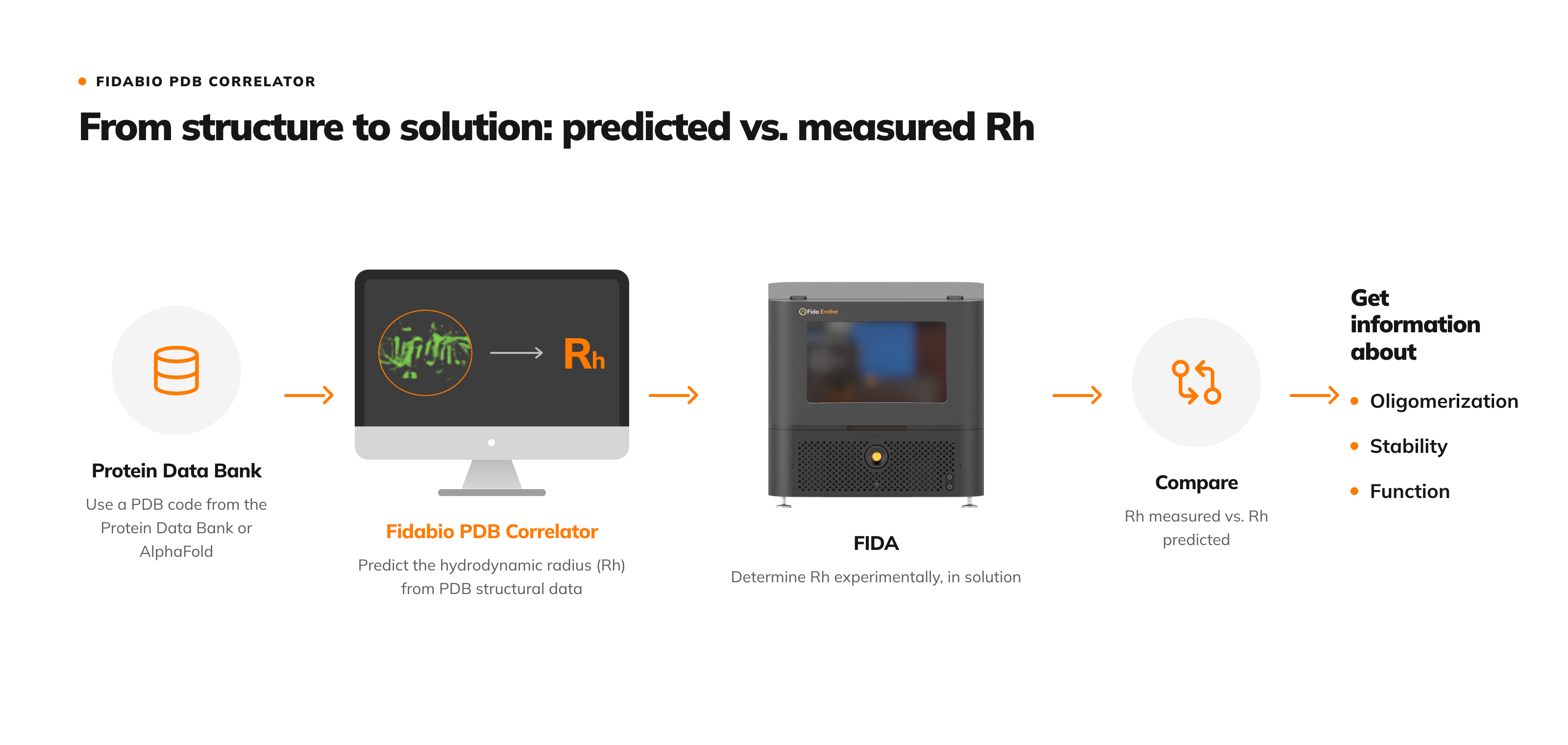
The PDB Correlator, a feature of the Fidabio software, predicts the hydrodynamic radius from the atomic coordinates in the structure file, together with the radius of gyration and the ratio between them, and compares the predicted hydrodynamic radius with the one FIDA measured.
How to use the PDB Correlator?
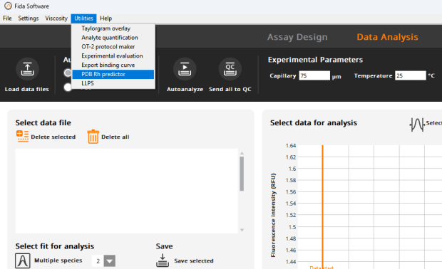
In the Fida software, open Utilities, then PDB Correlator.
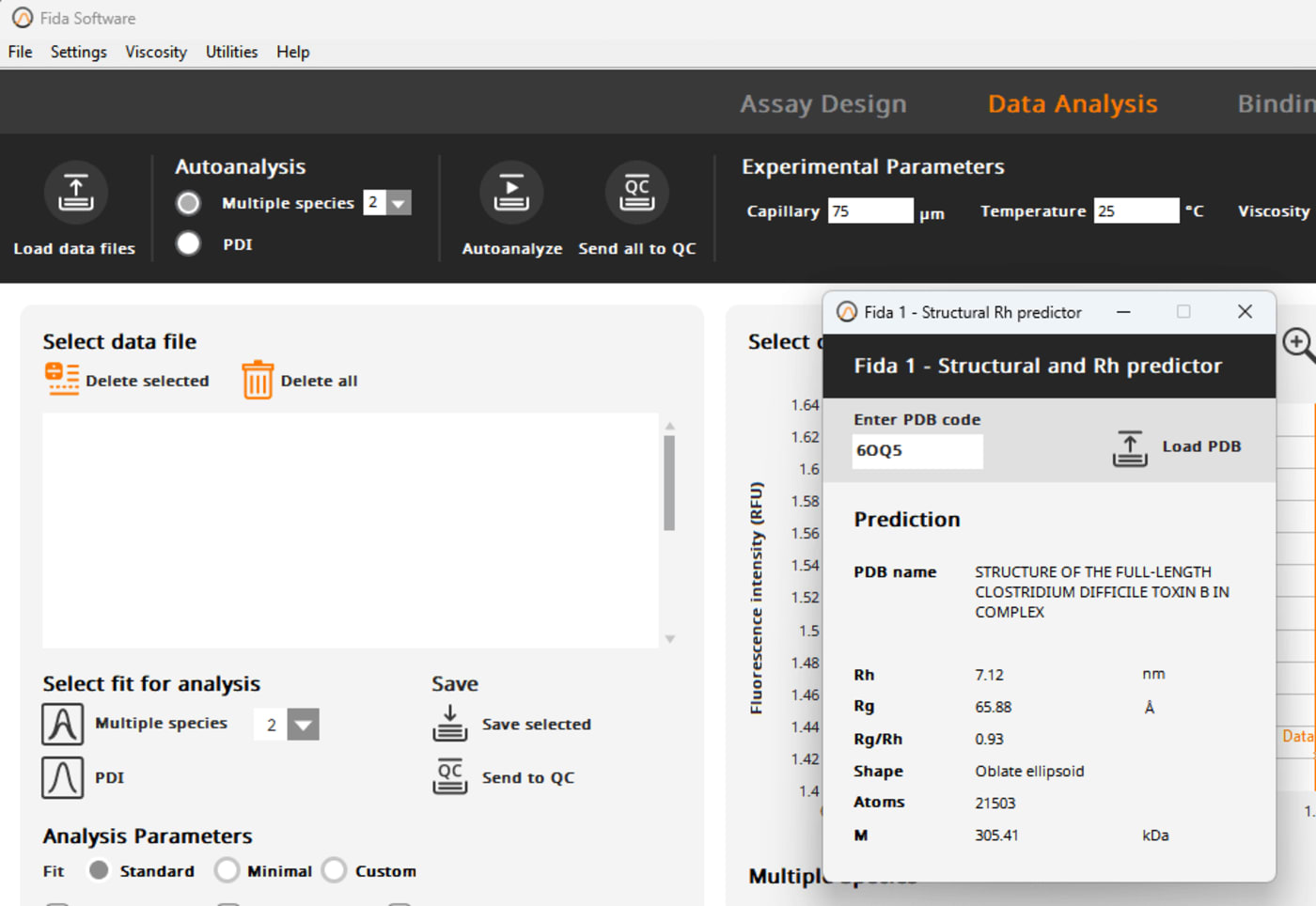
There are two ways for you to upload your PDB file. You can either:
- upload your own PDB file by pressing the "Load PDB", or
- fetch the data from the PDB by inserting the 4 digit PDB code for your structure.
The PDB correlator will then calculate for you the hydrodynamic radius (Rh), the radius of gyration (Rg), and the ratio between Rh and Rg. It will also tell you the name, shape, number of atoms, and molecular weight of your structure. You can also see this application note for more: See our Application Note about the PDB Correlator.
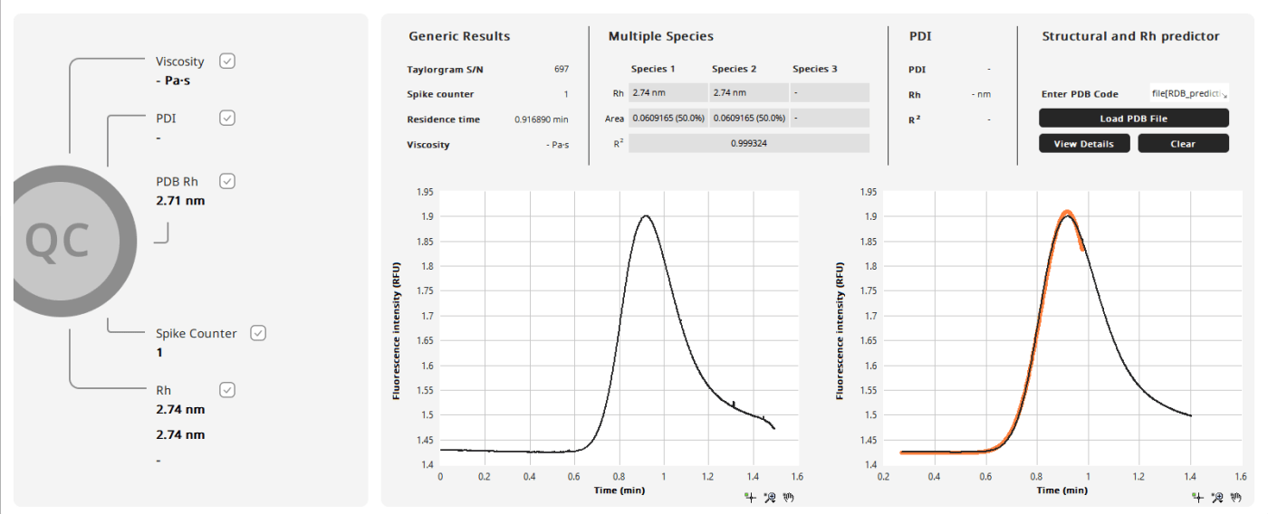
If the measured and predicted sizes agree, the oligomeric state of the protein is confirmed. If they differ, the protein may not be folded correctly, or it may form structures of higher order than 1:1. This makes the comparison a quality control step, for example to confirm that the sample is indeed what is expected.
How do you compare an AlphaFold model with the measured size of your protein?
AlphaFold generates a PDB file from the protein sequence, so the PDB Correlator works with an AlphaFold prediction in the same way as with a crystal structure. Either one gives the hydrodynamic radius to expect when the molecule or complex is measured with FIDA.
Why does the measured size of my protein differ from its crystal structure?
There are a few things to keep in mind when interpreting the data from the PDB correlator.First of all, it is important to keep in mind that proteins are dynamic molecules, and the structure of proteins in solutions can differ from their crystal structure
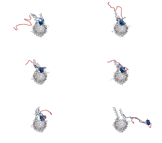
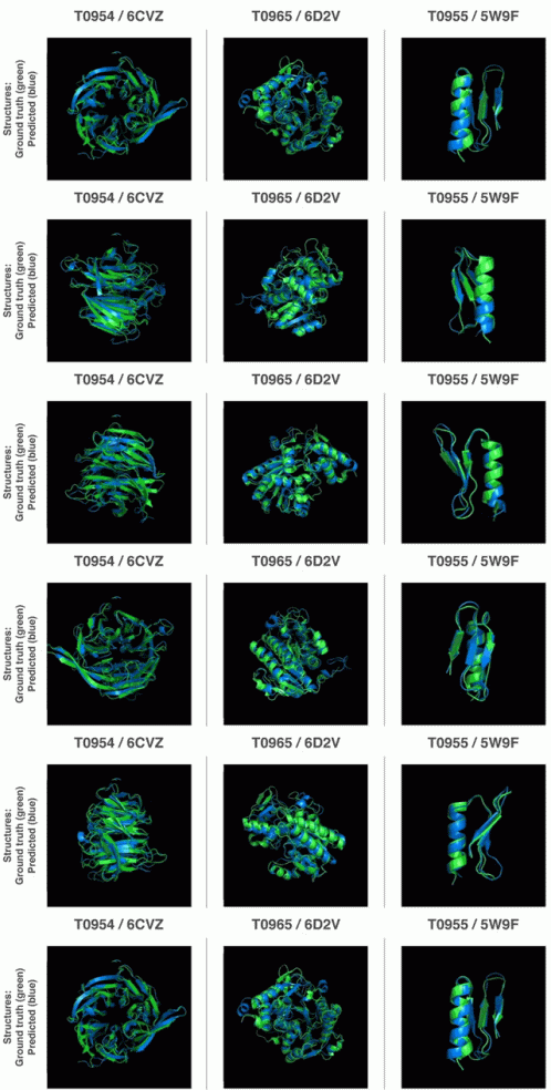
(GIF is from Heidarsson et al., 2022). In a crystal, proteins are packed into a lattice, which can introduce constraints and interactions not present in solution. These packing forces may distort flexible regions, stabilize certain conformations, or restrict motion. In solution, proteins are free to adopt a range of conformations influenced by solvent interactions, pH, ionic strength, and temperature. So, you might observe minor differences between the predicted hydrodynamic radius and the one you measure on the Fida instrument. Another thing to consider is that your protein might not be on the from you expect it to be. At your working concentration, your protein might form dimers or trimers, or even oligomerize further. This can, however, be investigated by building your dimer or oligomer in PyMOL and using the PDB correlator to predict the hydrodynamic radius of that molecule. See What is protein oligomerisation?
Frequently asked questions
What is the Protein Data Bank?
The Protein Data Bank (PDB) is “the single worldwide archive of structural data of biological macromolecules”, as Helen M. Berman and colleagues describe it in Nucleic Acids Research (2000). Each entry has its own PDB code.
What is a PDB code?
A PDB code, or PDB ID, is the unique four-character identifier of an entry in the Protein Data Bank. The first character is a number from 1 to 9, and the other three are letters or numbers, as defined by Protein Data Bank Japan; 4HHB, for example, is the crystal structure of human deoxyhaemoglobin (RCSB PDB). In the FIDA software, entering the PDB code fetches the structure directly from the Protein Data Bank.
What is AlphaFold?
AlphaFold is a neural network-based model from DeepMind that predicts the three-dimensional structure of a protein from its amino acid sequence alone. In the CASP14 structure prediction assessment it showed “accuracy competitive with experimental structures in a majority of cases”, as John Jumper and colleagues report in Nature (2021). Predicted structures are openly available in the AlphaFold Protein Structure Database, described by Mihaly Varadi and colleagues in Nucleic Acids Research (2022). Thanks to the PDB Correlator in the Fidabio software, you can now effortlessly check the size of your protein measured with FIDA against its AlphaFold structure, even when no crystal structure exists. Read this application note to see how.
What is the radius of gyration, and how does it differ from hydrodynamic radius?
The radius of gyration (Rg) is the mass-weighted root mean square distance of a molecule's mass elements from its centre of mass, and comes from a scattering measurement or from a structure. The hydrodynamic radius (Rh) is the effective radius a molecule has as it diffuses, including the water it carries. The ratio between them reflects shape rather than size. What is hydrodynamic radius? explains both in more detail.
Do you need a crystal structure to use the PDB Correlator?
No. The PDB Correlator works with any PDB file, including an AlphaFold prediction generated from the protein sequence.
Can you use a cryo-EM structure?
Yes. Measured size can be correlated with X-ray, cryo-EM and AlphaFold structures, as shown in the application note Linking Structure and Function.
Can the PDB Correlator predict the size of a dimer or complex?
Yes. Build the dimer or complex in PyMOL, then use the PDB Correlator to predict its hydrodynamic radius and compare it with the radius FIDA measures. The PDB Correlation course in the Sample Characterization & QC section of the Fidabio Learning Hub shows how.
Can you estimate the size of a protein complex before measuring it?
Yes. The PDB Correlator can estimate the hydrodynamic radius of the fully bound complex from its structure, so it does not need to be determined experimentally. When a binding curve does not reach a plateau at higher concentrations, entering and fixing this complex size helps the software find the top of the curve and improves the accuracy of the KD. The Data Interpretation course in the Affinity section of the Fidabio Learning Hub shows how.
Related resources
Want to see measured and predicted size side by side? The poster PDB Correlator: Linking Structure and Function compares them for β-lactoglobulin, human and bovine serum albumin, and lysozyme. For a one-page overview of how a PDB, cryo-EM or AlphaFold structure becomes a size you can check your sample against, take a look at the Fidabio PDB Correlator infographic.
Curious how decades of Protein Data Bank structures and AlphaFold predictions can be put to work on your own samples, in just a few clicks? Read PDB Correlator: how can it help you to establish a link between structure and functionality?
New to hydrodynamic radius? What is hydrodynamic radius? explains what the predicted and measured values mean, and Stokes-Einstein equation: symbols and SI units shows how a measured diffusion coefficient becomes a radius. The PDB Correlator readout sums up what the tool gives you in every analysis.
No structure yet? Get a quick estimate of your protein's size from its molecular weight with the hydrodynamic radius and molecular weight calculator.
Already a FIDA user? The full PDB Correlation course is waiting for you on the Fidabio Learning Hub.