What is protein stickiness?
Protein stickiness is the tendency of a molecule or sample component to adsorb non-specifically to surfaces and assay components, such as glassware, plastic labware, vials, well plates or a measurement capillary. It is an unspecific interaction between the protein and the surface, rather than binding to a defined site.
The difficulty with stickiness is that it is subtle and hard to quantify. There is usually no visual cue that it is happening, so it commonly goes undetected and is mistaken for something else.
What causes a protein to stick?
Stickiness arises from ordinary physical chemistry rather than from any specific binding site. Proteins are prone to it by nature, because many carry hydrophobic regions or charged side chains that interact with glass and plastic.
Common lab plastics such as polypropylene are hydrophobic, which can cause proteins to bind via hydrophobic patches. Electrostatic interaction operates wherever the protein and the surface carry opposite charges. Van der Waals forces are weak individually but become significant across a large contact area. Proteins may also unfold slightly at interfaces, which increases their tendency to stick. The presence of specific binding sites can contribute as well.
On a standard capillary the interaction is most likely between positive charges on the protein and the negative charge on glass. Fused silica, the material a measurement capillary is made from, carries an induced negative charge on its inner surface. A protein with a net positive charge, or one containing domains rich in positive charge, interacts electrostatically with that wall.
Why does stickiness matter?
Protein adsorbed onto a surface is no longer part of the sample, and three consequences follow.
Protein concentration is underestimated, because measured values may be lower than the actual ones. Reproducibility is poor, because stickiness is not always consistent, which leads to assay variability. And rare or precious material is lost, which can be critical in clinical or research settings with limited material.
There is also a consequence beyond the current experiment. A molecule that sticks to a FIDA capillary will also stick to other setups involving glass, so the behaviour carries into downstream biophysical analysis rather than being confined to one measurement.
How does stickiness reveal itself?
In Flow Induced Dispersion Analysis (FIDA), stickiness is visible in the raw signal rather than hidden inside a final number, and it appears in four recognisable patterns.
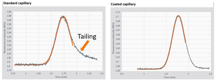
The first is a tail on the peak. An asymmetric Taylorgram, with a drag or tail effect on a standard capillary, indicates stickiness to the glass capillary. Measuring the same sample on a coated capillary resolves it, and the peak is seen as a symmetric gaussian.
The second is a loss of signal when the binding partner is added: the indicator alone behaves well, then fluorescence falls once the analyte is introduced, which indicates that the analyte is adsorbing to the capillary and taking the complex with it.
The third is a signal that fades over time, where repeated measurements of the same sample give progressively lower intensity or peak area, pointing to adsorption in the well plate or vial rather than in the capillary.

The fourth is an artificially late peak. In cases of extreme stickiness the Taylorgram can appear to shift to the right, as if there were an increase in viscosity. If the sample should have the viscosity of water, that cannot be true. The top of the Taylorgram is always at double the time of the indicator aggregates, and at 400 mbar, 25 degrees Celsius and the viscosity of water this is approximately 1.8 minutes.
Each pattern points to a different cause and therefore to a different remedy. How do you stop protein sticking to surfaces? works through them in order.
How stickiness is measured in FIDA
In FIDA the sample is carried through a fused silica capillary in free solution and its dispersion profile is recorded as it passes the detector. Surface interaction changes the shape of that profile, so stickiness is read from the measurement itself rather than inferred afterwards. In contrast to most biophysical techniques, which give a result without indicating whether adsorption occurred, FIDA returns stickiness as a readout.
The peak shape is the measurement. A protein that interacts with the capillary surface is sticky, and it is seen as a non-symmetrical Taylorgram, typically a skewed gaussian signal. Comparing the same sample on a standard capillary and on a coated one is diagnostic: if the profile is tailed on fused silica and symmetric on the coated capillary, the asymmetry came from the surface and not from heterogeneity in the sample.
Moderate stickiness does not prevent a measurement. A hydrodynamic radius can still be obtained from a partly distorted peak by fitting a defined fraction of it, for example the leading 51 percent, and reconstructing the remainder on the assumption of symmetry, although sizing is more reliable when stickiness has been minimised.
Because the sample is measured in free solution with nothing immobilised, stickiness is recorded alongside size, binding and aggregation in the same run, and is available as a quality parameter rather than as a separate experiment.
Related resources
Stickiness is one of the quality parameters returned by every FIDA run. The application note Assessment of Sample Quality with Every Measurement covers the full set, and the infographic Quality Control: 8 Parameters in 4 Minutes shows them at a glance. For worked examples across different protein systems, see the poster Applications of FIDA in Protein Science, and the case discussion Solving difficult cases at core facilities from the Biozentrum Basel.
Read next: How do you stop protein sticking to surfaces?, the Sample Stickiness readout and What is protein aggregation?